

Point of View: Reflections on Working with the Neck Down
Horse Anatomy vs “Anatomy”
The horse’s ability to carry itself is not limited to collection. It is a natural consequence of the proper mental and muscular development of the horse.
There are two types of approaches to horse anatomy: one that supports the belief that lowering the neck stretches the muscles of the horse’s upper and back muscles, and the actual anatomy of the horse that contradicts this view. Even if in their theoretical basis both approaches refer to the same horse, they are completely different.
One of the strong arguments for low neck and stretched muscles is that by stretching the head and nose forward, the horse increases the stretch in the upper neck muscles. A commonly used expression is “pulling over the reins”. The problem with this statement is that the muscles that move the head are not the same muscles that lower the neck. There are 21 pairs of muscles that move the head, including those responsible for pulling the nose forward. They do not lengthen the muscles of the upper neck at all. In fact, pulling the nose forward shortens the upper sections of the muscles of the upper neck.
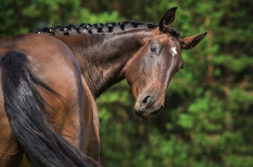
The two main muscles involved in lowering the neck are the lamellar and the semispinous. The patch-like muscle does not even have an attachment to the skull (attached laterally to the occiput). In addition to its primary function of resisting neck descent, the lateral attachment of the lamellar muscle to the superior end of the occiput allows the muscle to produce lateral flexion of the head and neck. In fact, the muscle does not lengthen at all when the horse’s nose is pulled forward.
Another main muscle of the upper part of the neck – the semispinous muscle of the head – still has an upper element that is attached to the top of the skull. However, in order to push the horse’s nose forward, the superior semispinalis muscle must pull the skull back, so the muscle does not lengthen, but, on the contrary, becomes shorter in concentric contraction. I am talking about a section of the muscle here because the semispinalis capis has numerous sections. The muscle has an internal tendon and 6 elements located below the central tendon, as well as 7 sections located above. Such a structure is convenient for the horse, as it allows for numerous movements, such as lateral (lateral) bending, rotation, etc. At the same time, such a structure contradicts theories of stretching, which proceed from the fact that the muscle lengthens as a whole. Due to the structure of the muscle sections, those sections that are at its base can perform an action that is completely different from that performed by the muscle section located at its upper part.
Most stretch theories tell us that the muscles in the lower neck, that is, the muscles below the cervical spine, pull the head and neck down, stretching the muscles in the upper neck. In fact, everything is quite the opposite. Lowering the neck is not provided by the muscles of the lower neck, as they cannot lengthen the muscles of the upper neck. Instead, the lowering of the neck is created by gravity. The horse’s head and neck make up about 10% of the horse’s total body mass, and gravity pulls the head and neck down.
The muscles of the upper neck resist gravity and therefore allow the head and neck to drop in the process of overcoming gravity. Some studies define upper neck muscle resistance as an isometric hold, meaning that the muscles contract without lengthening or shortening. Other studies define muscle action as eccentric, meaning that the muscle contracts during lengthening. These data must be reasonably interpreted. Active stretch or eccentric contraction is the most powerful type of muscle contraction. Such contractions can be 15 to 50 times stronger than a concentric contraction.
Stretch theories often use the term “relaxation”. In any position of the neck, the lamellar muscle is tensed as the forehoof produces a dynamic force that counteracts the acceleration of gravity created by dynamic loads. This tension, which occurs twice in one step, is part of the musculoskeletal mechanism at walk, trot and canter. If the neck were relaxed, then the neck and head would drop from each dynamic impact.
The main argument of stretching proponents is that the horse “stretches” naturally by dropping its head down after work. There is a strong ligament – the nuchal – connecting the cranio-thoracic spine with the skull. The nuchal ligament is not in tension when the horse’s neck is raised at the moment of alarm. It tenses up when the horse’s neck is lowered into a more horizontal position. The purpose of the nuchal ligament is to reduce the workload on the muscles of the upper neck. At a walk, it facilitates the work of the muscles of the upper part of the neck by 55%, at a trot and canter – by 32 – 36%. The horse does not stretch the neck – it simply facilitates the work of the muscles of the upper neck by placing the neck in a more horizontal position and thus using the passive resistance of the nuchal ligament.
Proponents of the low neck and stretched muscles theory make claims, but rarely explain their essence. And when they explain, they invent the anatomy of the horse, corresponding to their beliefs. I recently saw in print an excellent example of “comfortable” anatomy—the type of anatomy that is consistent with, but not even faintly related to, the physiology of the horse and its actual functioning: the sprain can extend as far as the hips.” This assertion is clearly false. The two main muscles of the back – the longissimus dorsi – are actually made up of several muscles that run in the same direction: the longissimus cervix, the head, the chest, the psoas, and so on.
The bundles of muscle fibers of the system of the longest muscle are attached by oblique muscles down and forward from the thoracic spine to the articular processes of the spinal column. The bundles connect approximately 3 to 5 vertebrae. The bundles of the multifidus muscle are oriented in the opposite direction, go obliquely, down and back, capturing about 3 vertebrae, all the while passing along the lumbothoracic spine. During movement, many circumstances occur when the bundles of muscle fibers of the lumbar-thoracic spine contract differently than the bundles of the lumbar spine.
Every time we publish a discussion about neck drop for educational purposes, we are the target of extremely nasty attacks from uneducated riders and trainers who want to believe in their stretch theories. In fact, it does not affect the anatomy of the horse in any way. We wish to provide further clarification if you wish to know more about the functional anatomy of the horse, in the light of cutting-edge modern research explaining it.
Jean Luc Corny (source).
Translation by Dina Maximova.
Here you can read stories about horses translated by Dina.
Neck lengthening. Fairy tales and reality
“People are willing to believe what they want to believe” (Gaius Julius Caesar)
Caesar’s idea applies to lowering the horse’s neck as well.
The horse’s neck stretched down as the horse tries to reach the snaffle is how we can describe the process of lowering the neck. But this concept goes against the concept of muscular work of the muscles of the upper neck when the neck is lowered. Building a well-conditioned dressage horse should be the ultimate goal of all training methods. However, proper body coordination is not the result of metaphors that involve stretching or telescoping the neck. These theories are fairy tales. They say that we want to create a horse whose body works effortlessly, but the theory of muscle work that its followers profess is far from reality.
Wishing the horse well is a wonderful emotion, but when the relationship with the horse is based on physical work and preparation for tournaments, a clear understanding of how the horse really works is a prerequisite.
The main muscles of the upper part of the neck are the lamellar and semispinalis muscles. Both are involved in raising the neck and resisting its lowering. The weight of the head and neck is approximately 10% of the horse’s body weight. This is a significant burden that falls under the influence of gravity. Without the resistance of the muscles of the upper neck, the horse’s head would sink down to the limit that is limited by the occipital ligament (this is what happens when the horse is under sedation, when the muscles of the upper neck no longer resist the force of gravity, and the occipital ligament works as a support). At the maximum test of elasticity, the occipital ligament holds the horse’s head a few centimeters off the ground. This is why horses spread their front legs when they are grazing so they can get to the grass.
The underlying architecture of the muscle, the absence of single contractions from origin to attachment, contradicts the theory that action at one end of a muscle will be transmitted to the other end. This simplistic thinking often reinforces theories that believe that lowering the neck lengthens the back muscles. Inside the muscle cells produce forces that are transmitted to other cells through connective tissues and so on. Muscle cells can create simultaneous forces acting in different directions. Connective tissues are also located within the muscle, allowing for multiple tasks to be performed simultaneously.
The distribution of connective tissue can contribute to the separation of its neuromuscular functions along the entire length of the muscle.
The morphology of the semispinalis muscle implies different functions of its dorsal and ventral regions. The muscle has a strong central tendon within its body which divides it into a dorsal region above the tendon and a ventral region below the tendon. This type of different architecture when dividing into dorsal and ventral parts of the same muscle is often seen when the function of the muscle cells is to increase tension in the central tendon. When the neck is lowered, the central tendon is stretched and the muscle cells work to increase the elastic resistance of the central tendon. It must be remembered that the horse’s head weighs about 10% of the horse’s body weight. As the neck descends, the upper neck muscles, tendons, and ligaments support the weight of the head against gravity.
A very simple experiment can be done that requires a large bucket of water, a broom and two elastic cords. The bucket of water is the horse’s head. The broom handle is a column of cervical vertebrae. One elastic cord is the muscles of the upper part of the neck. Another cord is used to attach a bucket of water to the tip of the broom handle. You attach one end of the elastic cord to the tip of the broom handle and hold the other end in your right or left hand. The elastic cord is the central tendon of the semispinalis muscle, and your arm works like the cells and connective tissues of the muscle. You block the base of the broom with your foot and pull on the elastic cord to raise the top end of the broom, which has a bucket of water hanging from it. By doing this, your arm works the way the pectoralis dorsi muscles of the horse’s neck work, as well as the dorsal element of the semispinalis muscle when they lift the horse’s neck. Then you lower the horse’s head, which in the experiment is a water bucket. According to the “stretch” theory, your arm should stretch. Of course, this is not true. You are pulling hard on the elastic cord, otherwise the bucket will fall down. In this way, you are doing the work of the muscles of the horse’s upper neck. The lower you lower the bucket of water, the more it pulls the central tendon, the elastic cord, and the more actively the muscle cells, which in the experiment are represented by the muscles of your arm, work.
The idea that the horse’s neck is pulled out of the shoulders and stretched and thus we achieve the desired result is wishful thinking. Even as a metaphor, this thought evokes completely false ideas. The problem is that the horse is not a fictitious model. Physical activity is the work of muscles, tendons, ligaments, fascia and nerve circuits. If the body coordination matches the rider’s imagination but is not related to the physiology of the horse, the horse will perform beyond its potential until lameness puts an end to everything. Lameness is not the only expression of physical pain. Anticipation of a given movement or (more generally) anticipation of entering the battlefield, restlessness, nervousness, gait abnormalities, frustration, anger, lock-in, and many other behaviors are manifestations of pain.
The question may be asked: but why, then, does the horse lower its neck on its own after work? The answer is simple. The horse facilitates the work of the muscles of the upper part of the neck, using the elastic resistance of the occipital ligament. The occipital ligament is attached to the first to fourth thoracic vertebrae. On the other hand, the funicular element of the occipital ligament is attached to the skull. The occipital ligament is not under tension when the neck is held high, but it is actually under tension when the horse lowers the neck. The occipital ligament, which can be compared to a strong elastic cord, lengthens to assist the muscles of the upper neck in their work of supporting the head and neck. The occipital ligament replaces 55% or more of the work of the upper neck muscles in the walk. At the trot and canter, it replaces 32 to 34% of the work of the upper neck muscles. As the horse lowers the neck, tension in the occipital ligament increases and the muscles in the upper neck become less active. There is no stretch; it’s just easing the work of the muscles of the muscles of the upper part of the neck.
The idea that lowering the neck increases the range of motion of the horse’s lumbar and thoracic spine is also inaccurate. The loss and increase in mobility of the vertebrae during the lowering of the neck were measured. Five subjects took part in the experiment. All five had vertebral mobility between T6 and T9, with the neck down. T6 and T9 are the front of the withers. This increase in mobility is mainly due to the fact that the supraspinatus ligament, which is a continuation of the occipital ligament and lies above the processes of the spine, still retains some elasticity until T9. One in five subjects lost mobility between T9 and T14 with the neck down. In the remaining four subjects, the mobility of the vertebrae in the same area did not increase when the neck was lowered. One of the subjects lost vertebral mobility between T14 and T18, with the neck down. In other subjects, mobility in this particular area did not increase with the neck down. All subjects lost vertebral mobility in the lumbar vertebrae. Five subjects had increased mobility in the lumbosacral region. This is the reason why the observations of researchers who are not well versed in this matter lead them to believe that the lumbar vertebrae flex when the neck is lowered.
In the XNUMXth century, the Prussian cavalry contributed to the fact that during the ride the riders lifted the horse’s neck completely. The idea behind the theory was to improve the balance of the horse. The experiment lasted several decades, and then completely stopped. My guess is that injuries have reached epidemic levels and even the greatest proponents of this idea have had to rethink it. In contrast, Paul Plinzner, who was a master horseman in the Emperor’s court, lowered and excessively bent the necks of his horses. It was the end of the 19th century. Since then, the nape of the horse has remained either the highest or lowest point of the neck. Whatever the fashion, horsemen constantly vacillated between the two positions for lack of evidence that either was correct. François Boche lifted his neck to new heights. Jacques Licard later emphasized neck stretching. The French author relied on stretching and mentioned the lowering of the neck as its lengthening. In fact, technically, lowering the neck is a flexion. The neck lift is an extension. More recently, Harry Bolt warned against dropping the neck very low, and so on. The common denominator of these conflicting theories is that they are based on a lack of science and a highly developed imagination. Another similarity is that while they promote diametrically opposed approaches, both theories talk about connecting the horse’s back and hindquarters.
The truth is that the effects associated with lowering the neck cannot be achieved by acting on the neck. The position of the neck is the result of suitable, most rational work, the result of the exact coordination of the horse’s body, starting with the slowing and driving activity of the hind legs and continuing with the ability of the muscles of the back to convert the impulse created by the hind legs into horizontal forces, forward movement, and vertical forces, resistance to the force of gravity, and therefore balance control. Proper spinal mechanics allows the forelegs to propel the horse’s body up and forward. The horse then places the neck in the correct position and uses it to further improve and control the balance, quality and accuracy of limb kinematics. The idea that such effective coordination could be the result of lowering the neck is a fantasy. The problem is that fiction does not effectively prepare the horse’s body for competition.
Translation by Valeria Smirnova (a source).


You May Also Like

Working with a shankel – we figure out how to do it right
Where is the horse looking?
