What is the heart of amphibians: a detailed description and characteristics
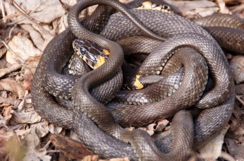
Amphibians belong to the class of four-legged vertebrates, in total this class includes about six thousand seven hundred species of animals, including frogs, salamanders and newts. This class is considered to be rare. There are twenty-eight species in Russia and two hundred and forty-seven species in Madagascar.
Amphibians belong to terrestrial primitive vertebrates, they occupy an intermediate position between aquatic and terrestrial vertebrates, because most species reproduce and develop in the aquatic environment, and individuals that have matured begin to live on land.
Amphibians have lungs, which they breathe, the blood circulation consists of two circles, and the heart is three-chambered. Blood in amphibians is divided into venous and arterial. The movement of amphibians occurs with the help of five-fingered limbs, and they have spherical joints. The spine and skull are movably articulated. The palatine square cartilage fuses with the autostyle, and the himandibular becomes the auditory ossicle. Hearing in amphibians is more perfect than in fish: in addition to the inner ear, there is also a middle ear. The eyes have adapted to see well at different distances.
On land, amphibians are not fully adapted to live – this can be seen in all organs. The temperature of amphibians depends on the humidity and temperature of their environment. Their ability to navigate and move on land is limited.
Contents
Circulation and circulatory system
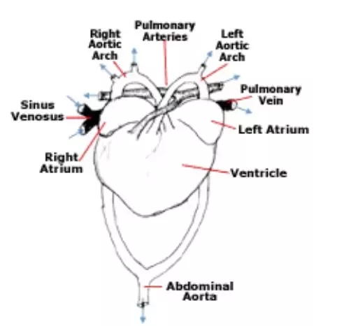
Amphibians have a three-chambered heart, it consists of a ventricle and atria in the amount of two pieces. In caudate and legless, the right and left atria are not completely separated. Anurans have a complete septum between the atria, but amphibians have one common opening that connects the ventricle to both atria. In addition, in the heart of amphibians there is a venous sinus, which receives venous blood and communicates with the right atrium. The arterial cone adjoins the heart, blood is poured into it from the ventricle.
The conus arteriosus has spiral valve, which distributes blood into three pairs of vessels. The heart index is the ratio of heart mass to percentage of body weight, it depends on how active the animal is. For example, grass and green frogs move very little and have a heart rate of less than half a percent. And the active, ground toad has almost one percent.
In amphibian larvae, the blood circulation has one circle, their blood supply system is similar to fish: one atrium in the heart and ventricle, there is an arterial cone branching into 4 pairs of gill arteries. The first three arteries split into capillaries in the external and internal gills, and the branchial capillaries merge in the branchial arteries. The artery that carries out the first branchial arch splits into carotid arteries, which supply the head with blood.
gill arteries
Merging the second and third efferent branchial arteries with the right and left aortic roots and their connection occurs in the dorsal aorta. The last pair of branchial arteries does not split into capillaries, because on the fourth arch into the internal and external gills, the aorta of the back flows into the roots. The development and formation of the lungs is accompanied by circulatory restructuring.
The atrium is divided by a longitudinal septum into left and right, making the heart three-chambered. The network of capillaries is reduced and turns into carotid arteries, and the roots of the dorsal aorta originate from the second pairs, the caudates retain the third pair, while the fourth pair turns into skin-pulmonary arteries. The circulatory peripheral system is also transformed and acquires an intermediate character between the terrestrial scheme and the water one. The largest restructuring occurs in amphibian anurans.
Adult amphibians have a three-chambered heart: one ventricle and atria in the amount of two pieces. The venous thin-walled sinus adjoins the atrium on the right side, and the arterial cone departs from the ventricle. It can be concluded that the heart has five sections. There is a common opening, due to which both atria open into the ventricle. The atrioventricular valves are also located there, they do not allow blood to penetrate back into the atrium when the ventricle contracts.
There is a formation of a number of chambers that communicate with each other due to the muscular outgrowths of the ventricular walls – this does not allow the blood to mix. The arterial cone departs from the right ventricle, and the spiral cone is located inside it. From this cone arterial arches begin to depart in the amount of three pairs, at first the vessels have a common membrane.
Left and right pulmonary arteries move away from the cone first. Then the roots of the aorta begin to depart. Two branchial arches separate two arteries: subclavian and occipital-vertebral, they supply blood to the forelimbs and muscles of the body, and merge in the dorsal aorta under the spinal column. The dorsal aorta separates the powerful enteromesenteric artery (this artery supplies the digestive tube with blood). As for other branches, the blood flows through the dorsal aorta to the hind limbs and to other organs.
Carotid arteries
The carotid arteries are the last to depart from the arterial cone and divided into internal and external arteries. The venous blood from the hind limbs and the part of the body located behind is collected by the sciatic and femoral veins, which merge into the renal portal veins and break up into capillaries in the kidneys, that is, the renal portal system is formed. The veins depart from the left and right femoral veins and merge into the unpaired abdominal vein, which goes to the liver along the abdominal wall, so it breaks up into capillaries.
In the portal vein of the liver, blood is collected from the veins of all parts of the stomach and intestines, in the liver it breaks up into capillaries. There is a confluence of the renal capillaries into the veins, which are efferent and flow into the posterior unpaired vena cava, and the veins extending from the genital glands also flow there. The posterior vena cava passes through the liver, but the blood that it contains does not enter the liver, small veins from the liver flow into it, and it, in turn, flows into the venous sinus. All caudate amphibians and some anurans retain cardinal posterior veins, which flow into the anterior vena cava.
arterial blood, which is oxidized in the skin, is collected in a large cutaneous vein, and the cutaneous vein, in turn, carries venous blood into the subclavian vein directly from the brachial vein. The subclavian veins merge with the internal and external jugular veins into the left anterior vena cava, which empty into the venous sinus. Blood from there begins to flow into the atrium on the right side. In the pulmonary veins, arterial blood is collected from the lungs, and the veins flow into the atrium on the left side.
Arterial blood and atria
When breathing is pulmonary, mixed blood begins to collect in the atrium on the right side: it consists of venous and arterial blood, venous blood comes from all departments through the vena cava, and arterial blood comes through the veins of the skin. arterial blood fills the atrium on the left side, blood comes from the lungs. When a simultaneous contraction of the atria occurs, blood enters the ventricle, the growths of the walls of the stomach do not allow the blood to mix: venous blood predominates in the right ventricle, and arterial blood predominates in the left ventricle.
An arterial cone departs from the ventricle on the right side, so when the ventricle contracts into the cone, venous blood first enters, which fills the skin pulmonary arteries. If the ventricle continues to contract in the arterial cone, pressure begins to increase, the spiral valve begins to move and opens the openings of the aortic arches, in them mixed blood rushes from the center of the ventricle. With full contraction of the ventricle, arterial blood from the left half enters the cone.
It will not be able to pass into the arched aorta and pulmonary cutaneous arteries, because they already have blood, which with a strong pressure shifts the spiral valve, opening the mouths of the carotid arteries, arterial blood will flow there, which will be sent to the head. If pulmonary respiration is turned off for a long time, for example, during wintering under water, more venous blood will flow into the head.
Oxygen enters the brain in a smaller amount, because there is a general decrease in the work of metabolism and the animal falls into a stupor. In amphibians that belong to the caudate, a hole often remains between both atria, and the spiral valve of the arterial cone is poorly developed. Accordingly, the most mixed blood enters the arterial arches than in tailless amphibians.
Although amphibians have blood circulation goes in two circles, due to the fact that the ventricle is one, it does not allow them to completely separate. The structure of such a system is directly related to the respiratory organs, which have a dual structure and correspond to the lifestyle that amphibians lead. This makes it possible to live both on land and in water to spend a lot of time.
Red bone marrow
The red bone marrow of tubular bones begins to appear in amphibians. The amount of total blood is up to seven percent of the total weight of an amphibian, and hemoglobin varies from two to ten percent or up to five grams per kilogram of mass, the oxygen capacity in the blood varies from two and a half to thirteen percent, these figures are higher compared to fish.
Amphibians have large red blood cells, but there are few of them: from twenty to seven hundred and thirty thousand per cubic millimeter of blood. The blood count of larvae is lower than that of adults. In amphibians, just like in fish, blood sugar levels fluctuate with the seasons. It shows the highest values in fish, and in amphibians, caudates from ten to sixty percent, while in anurans from forty to eighty percent.
When summer ends, there is a strong increase in carbohydrates in the blood, in preparation for wintering, because carbohydrates accumulate in the muscles and liver, as well as in spring, when the breeding season begins and carbohydrates enter the blood. Amphibians have a mechanism of hormonal regulation of carbohydrate metabolism, although it is imperfect.
Three orders of amphibians
Amphibians are divided into the following divisions:
- Amphibians tailless. This detachment contains about one thousand eight hundred species that have adapted and move on land, jumping on their hind limbs, which are elongated. This order includes toads, frogs, toads, and the like. There are tailless on all continents, the only exception is Antarctica. These include: real toads, tree frogs, round-tongued, real frogs, rhinoderms, whistlers and spadefoot.
- Amphibians caudate. They are the most primitive. There are about two hundred and eighty species of them all. All kinds of newts and salamanders belong to them, they live in the northern hemisphere. This includes the protea family, lungless salamanders, true salamanders, and salamanders.
- Amphibious legless. There are approximately fifty-five thousand species, most of them live underground. These amphibians are quite ancient, having survived to our times due to the fact that they managed to adapt to a burrowing lifestyle.
Amphibian arteries are of the following types:
- Carotid arteries supply the head with arterial blood.
- Skin-pulmonary arteries – carry venous blood to the skin and lungs.
- The aortic arches carry blood that is mixed to the remaining organs.
Amphibians are predators, salivary glands, which are well developed, their secret moisturizes:
- language
- food and mouth.
Amphibians arose in the middle or lower Devonian, that is about three hundred million years ago. Fish are their ancestors, they have lungs and have paired fins from which, quite possibly, five-fingered limbs were developed. Ancient lobe-finned fish just meet these requirements. They have lungs, and in the skeleton of the fins, elements similar to parts of the skeleton of a five-fingered terrestrial limb are clearly visible. Also, the fact that amphibians descended from ancient lobe-finned fish is indicated by the strong similarity of the integumentary bones of the skull, similar to the skulls of amphibians of the Paleozoic period.
Lower and upper ribs were also present in lobe-finned and amphibians. However, lungfish, which had lungs, were very different from amphibians. Thus, the features of locomotion and respiration, which provided the opportunity to go on land in the ancestors of amphibians, appeared even when they were just aquatic vertebrates.
The reason that served as the basis for the emergence of these adaptations was, apparently, the peculiar regime of reservoirs with fresh water, and some species of lobe-finned fish lived in them. This could be periodic drying or lack of oxygen. The most leading biological factor that became decisive in the break of the ancestors with the reservoir and their fixation on land is the new food that they found in their new habitat.
Respiratory organs in amphibians
Amphibians have the following respiratory organs:
- The lungs are the respiratory organs.
- Gills. They are present in tadpoles and some other inhabitants of the water element.
- Organs of additional respiration in the form of skin and mucous lining of the oropharyngeal cavity.
In amphibians, the lungs are presented in the form of paired bags, hollow inside. They have walls that are very thin in thickness, and inside there is a slightly developed cell structure. However, amphibians have small lungs. For example, in frogs, the ratio of the surface of the lungs to the skin is measured at a ratio of two to three, compared with mammals, in which this ratio is fifty, and sometimes a hundred times greater in favor of the lungs.
With the transformation of the respiratory system in amphibians, change in breathing mechanism. Amphibians still have a rather primitive forced type of breathing. Air is drawn into the oral cavity, for this the nostrils open and the bottom of the oral cavity descends. Then the nostrils are closed with valves, and the floor of the mouth rises due to which air enters the lungs.
How is the nervous system in amphibians
In amphibians, the brain weighs more than in fish. If we take the percentage of brain weight and mass, then in modern fish that have cartilage, the figure will be 0,06–0,44%, in bone fish 0,02–0,94%, in amphibians tailed 0,29–0,36 %, in tailless amphibians 0,50–0,73%.
The forebrain of amphibians is more developed than that of fish; there was a complete division into two hemispheres. Also, development is expressed in the content of a larger number of nerve cells.
The brain is made up of five sections:
- Relatively large forebrain, which is divided into two hemispheres and contains olfactory lobes.
- Well developed diencephalon.
- Underdeveloped cerebellum. This is due to the fact that the movement of amphibians is monotonous and uncomplicated.
- The center of the circulatory, digestive and respiratory systems is the medulla oblongata.
- Vision and skeletal muscle tone are controlled by the midbrain.
The lifestyle of amphibians
The lifestyle that amphibians lead is directly related to their physiology and structure. The respiratory organs are imperfect in structure – this applies to the lungs, primarily because of this, an imprint is left on other organ systems. Moisture constantly evaporates from the skin, which makes amphibians dependent on the presence of moisture in the environment. The temperature of the environment in which amphibians live is also very important, because they do not have warm-bloodedness.
Representatives of this class have a different lifestyle, so there is a difference in structure. The diversity and abundance of amphibians is especially high in the tropics, where there is high humidity and almost always the air temperature is high.
The closer to the pole, the less amphibian species become. There are very few amphibians in the dry and cold regions of the planet. There are no amphibians where there are no reservoirs, even temporary ones, because eggs can often develop only in water. There are no amphibians in salt water bodies, their skin does not maintain osmotic pressure and hypertonic environment.
Eggs do not develop in salt water reservoirs. Amphibians are divided into the following groups according to the nature of the habitat:
- water,
- terrestrial.
Terrestrial can go far from water bodies, if this is not the breeding season. But aquatic, on the contrary, spend their entire life in water, or very close to water. In caudates, aquatic forms predominate, some species of anurans can also belong to them, in Russia, for example, these are pond or lake frogs.
Arboreal amphibians widely distributed among terrestrial, for example, copepod frogs and tree frogs. Some terrestrial amphibians lead a burrowing lifestyle, for example, some are tailless, and almost all are legless. In land dwellers, as a rule, the lungs are better developed, and the skin is less involved in the respiratory process. Due to this, they are less dependent on the humidity of the environment in which they live.
Amphibians are engaged in useful activities that fluctuate from year to year, it depends on their number. It is different at certain stages, at certain times and under certain weather conditions. Amphibians, more than birds, destroy insects that have a bad taste and smell, as well as insects with a protective color. When almost all insectivorous birds sleep, amphibians hunt.
Scientists have long paid attention to the fact that amphibians are of great benefit as insect exterminators in vegetable gardens and orchards. Gardeners in Holland, Hungary and England specially brought toads from different countries, releasing them into greenhouses and gardens. In the mid-thirties, about one hundred and fifty species of aga toads were exported from the Antilles and Hawaiian Islands. They began to multiply and more than a million toads were released onto the sugar cane plantation, the results exceeded all expectations.
Vision and hearing of amphibians

Amphibian eyes protect against clogging and drying out movable lower and upper eyelids, as well as the nictitating membrane. The cornea became convex and the lens lenticular. Basically, amphibians see objects that move.
As for the hearing organs, the auditory ossicle and the middle ear appeared. This appearance is due to the fact that it became necessary to better perceive sound vibrations, because the air medium has a higher density than water.


You May Also Like

Types of domestic turtles: description, lifestyle, diet and requirements for keeping at home
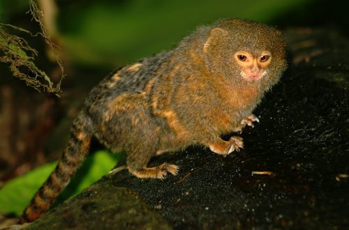
The smallest member of the primate group is the marmoset monkey.