Hip dysplasia in dogs
Hip dysplasia in dogs underdevelopment of the acetabulum of the hip joint, this results in a discrepancy between the shape of the femoral head and the acetabulum, and friction occurs and, as a result, erasure and destruction of the articular tissue (osteoarthritis), which, in turn, leads to severe disorders in the musculoskeletal system. Nowadays, this is a widespread disease that is typical for dogs of large breeds (Labrador, German Shepherd, Boxer, Newfoundland, etc.), but small breeds are also often susceptible to this disease. The basis of hip dysplasia in dogs is a genetic predisposition, but malnutrition and lack of exercise during the period of active growth makes a significant contribution to the development of this disease. A genetic predisposition does not always lead to the development of dysplasia. Dysplasia can manifest itself at any age, but most often structural changes in the joint begin to appear at 5-12 months, but the degree of dysplasia is established at the age of 1 year, in giant breeds – at 1.5 years.
Contents
The main symptoms of hip dysplasia in dogs
- Morbidity
- Lameness
- Increased fatigue while driving
- Stiffness of movements
- Unsteadiness of the hind limbs
- After resting, the animal gets up hard
- X-shaped posture of limbs
- In advanced cases, it can lead to immobilization
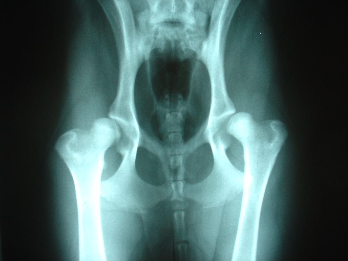
Diagnosis of hip dysplasia in dogs
The main and most revealing method of research is x-ray examination. It has the following requirements:
- The picture is taken in the position of the animal lying on its back with the pelvic limbs extended parallel to each other.
- Be sure to use sedatives or muscle relaxants.
- On the x-ray, indicate the pedigree number, breed, date of the x-ray, and indicate the right and left sides.
- An identification number is affixed to the radiograph using a digital X-ray stencil.
- The x-ray must have a clear image.
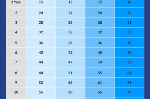
Degrees of hip dysplasia in dogs
Grade A – no signs of dysplasiaGrade B – joint alignment is close to normalGrade C – mild manifestation of hip dysplasiaGrade D – moderate manifestation of hip dysplasiaGrade E – severe manifestation of hip dysplasia.
Classification of lesions according to Schnell
Normal hip joint:
- joint angle 105о and more
- the anterior edge of the acetabulum to the outer end has a uniform convexity
- femoral neck without deposits
- the joint space is located in the cavity.
Hip dysplasia I degree:
- joint angle 100 – 105о
- thickening of the anterior edge of the acetabulum
- weak layering on the femoral neck
- low fixation of the femoral head.
Hip dysplasia II degree:
- joint angle less than 100о
- distinct layers on the neck of the femur
- low fixation of the femoral head.
Hip dysplasia III degree:
- flattened acetabulum
- signs of osteoarthritis
- subluxation of the femoral head.
Hip dysplasia IV degree:
- changes, as in grade III dysplasia.
- dislocation of the femoral head.
Treatment of hip dysplasia in dogs
Depending on the stage of hip dysplasia in dogs, the veterinarian selects the treatment. There are two methods: therapeutic and surgical. The therapeutic method is applied at grade (B, C). It includes chondroprotectors, painkillers and anti-inflammatory drugs, minimization of physical activity, the introduction of feed additives into the diet, and diet therapy in case of excess body weight. In more severe degrees (D, E) or in the absence of a positive response to therapeutic treatment, a surgical method is resorted to. Types of surgical interventions:
- Resection arthroplasty is used for animals weighing less than 30 kg in the presence of signs of osteoarthritis. It consists in removing the femoral head to eliminate injury to the acetabulum and reduce pain.
- Triple pelvic osteotomy – the basic principle is to change the angle of the acetabulum in order to completely cover the femoral head. It is forbidden to perform the operation in the presence of osteoarthritis and osteophytes. Its main goal is to save the legs, as well as relieve the animal from pain.
- Interventral osteotomy – used for valgus deformity of the thigh. In this case, dysplasia develops due to an incorrect inclination of the cervical-diaphyseal angle of the femur, and not due to underdevelopment of the acetabulum. The principle of treatment is to change the angle of the femoral neck, thereby preventing the destruction of the edge of the acetabulum.
Prevention of hip dysplasia in dogs
It is important to remember that any disease is easier to prevent than to cure. The main principles for the prevention of dysplasia are balanced feeding, normalization of physical activity and maintaining the normal weight of the animal.


You May Also Like
What to expect if a three-legged cat or three-legged dog appears in the house
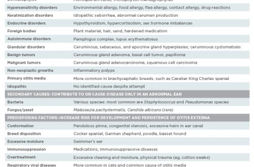
Otitis in dogs – causes, symptoms, types, treatment