Eye diseases in dogs and cats

One of the most common reasons for visiting a veterinarian is various eye diseases. Consider some diseases, for a timely visit to the veterinarian.
Symptoms of eye diseases
The main signs of problems with the eyes and periocular structures include:
- Epiphora – excessive lacrimation.
- Blepharospasm is the squinting of one or both eyes.
- Purulent discharge from the eyes.
- Photophobia.
- Purulent discharge from the eyes.
- Itching of the eyelids.
Most Common Eye and Eye Diseases in Dogs and Cats
Common eye conditions include:
- Inversion and eversion of the eyelids. The most common pathology of the lower eyelid. Eversion is dangerous due to infection, the possibility of developing dry keratoconjunctivitis. When twisting, the cornea is mechanically injured by the eyelashes, which can lead to an ulcer. Surgical treatment. The problem is usually congenital. It is more common in Maine Coons, Sharpei, Bulldogs, Central Asian Shepherd Dogs.
- Blepharitis of the eyelids. Eyelids can become inflamed both due to infection, mechanical trauma, and due to an allergic reaction. The disease requires emergency treatment, as it is often combined with other pathological conditions of the eye. Therapy depends on the cause of inflammation – sometimes antibiotics, drugs against microbes, antiallergic drugs are prescribed.
- Tumors of the eyelids. They can occur both on the upper and lower eyelids, and on the third. Diagnosis requires a fine needle biopsy of the neoplasm followed by cytological examination. This is followed by surgical excision or chemotherapy.
- Dry eye syndrome. It can develop due to many pathologies. Chronic disease, manifested by a decrease in the production of lacrimal fluid and accompanied by corneal-conjunctival xerosis (drying and keratinization of the epithelium).
Pathology occurs in most dogs, less often in cats. Normally, the tear film covers the entire surface of the cornea and conjunctiva. With insufficient tears, this film is torn, its protective function is lost. Dry keratoconjunctivitis or dry eye syndrome brings severe discomfort to the animal. It begins gradually, itching, burning, heaviness of the eyelids, a feeling of a foreign body in the eye. In the initial stages, there is reddening of the conjunctiva, copious discharge from the eyes. As the disease progresses, dryness of the conjunctiva develops, the animal squints and scratches the eyes, and abundant purulent and mucous discharges appear. In advanced cases, the cornea of the eye is affected, erosion may appear, and then corneal ulcers. In chronic course, there is a deposition of dark pigment in the cornea and the development of pigmentary keratitis. Treatment of dry keratoconjunctivitis is long, sometimes lifelong, antibiotics and artificial tears are used.
- Conjunctivitis is an inflammatory disease that is characterized by swelling, redness, soreness of the conjunctiva, purulent and clear discharge. It can occur for many reasons. As a rule, antibiotic drops are prescribed.
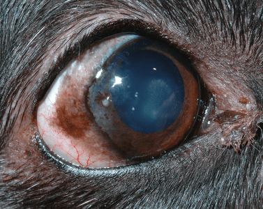
- Keratitis. Keratitis is called inflammation of the cornea, associated with a violation of its luster and transparency. It causes discomfort in the animal, reduces the quality of life and is fraught with dangerous complications. In most cases, after keratitis, persistent opacities remain on the cornea due to scarring of the surface layers. Negative consequences can be avoided with timely access to a doctor.
- Cataract. Cataract. It can be both senile and caused by some other diseases, such as diabetes. There are no drops that can help in the treatment. The only way is surgery, lens replacement.
- Glaucoma is an increase in intraocular pressure. May develop due to, for example, increased intracranial pressure or chronic renal failure. A feature of this disease is atrophy of the optic nerve. Increases in size and hardens the eyeball due to increased eye pressure; the mucous membrane of the eye swells; the cornea of the eye becomes cloudy and loses sensitivity. For treatment, the root cause is taken under control and special therapeutic drops are prescribed; in advanced cases, surgical methods are used.
- Uveitis is inflammation of the vasculature of the eye. It can be manifested by clouding of the cornea, redness of the sclera. The causes may be trauma, infectious diseases, have an idiopathic character. Drops are used for treatment, however, there may be no effect if the disease is caused, for example, by severe incurable infectious diseases: leukemia, immunodeficiency, infectious peritonitis of cats.
- Luxation (dislocation) of the lens. Pathology of the eye associated with displacement (luxation, dislocation, dislocation) of the lens from its normal anatomical position.
This pathology occurs more often in dogs than in cats. The disease is inherited and is called primary lens luxation (Primiry Lens Luxation – PLL). Both eyes are affected. It most often occurs at the age of 5 years. Secondary lens luxation is the result of a concomitant pathology in the eye that causes lens displacement (cataract, glaucoma, etc.). So, in cats, mainly secondary luxation of the lens occurs. The reasons for the development of lens luxation in dogs and cats are associated with weakness and rupture of the ligaments that hold the lens around the entire circumference in a strict position. As a result of tearing of these ligaments, the lens is displaced in different directions: into the anterior chamber, into the vitreous body, infringement in the pupil’s opening. Treatment is medical or surgical.
- Erosions and ulcers of the cornea. They arise as a complication of other diseases, for example, of an infectious or traumatic nature. To confirm the diagnosis, a test with fluorescein is performed. When the diagnosis is confirmed, a protective collar is put on the animal and a drug regimen is prescribed: an antibiotic, an anesthetic, a drug to restore the cornea.
- Prolapse of the third century. A common pathology is prolapse of the lacrimal gland tissue from the inner corner of the eye. Previously, the eyelid was simply removed, but this led to the development of dry eye syndrome. To date, mechanical reduction is successfully carried out, sometimes suturing is required for fixation.
- Eye injury. Animals are more likely to suffer during active games with each other or children, dogs with bulging eyes. Also, foreign bodies that have fallen into the conjunctival sac can injure the eyeball. Damage is usually accompanied by unilateral lacrimation and blepharospasm. The veterinarian excludes other diseases and prescribes symptomatic therapy aimed at pain relief, restoration of eye structures and prevention of infections.
- Blepharospasm is a symptom that can be a sign of various diseases of the eyelids and eyes. Neurological pathology in which the dog cannot control the work of the eyelids. The circular muscles of the eyes are involuntarily accelerated contraction. Because of this, the animal is not able to fully open its eyes and navigate in space. This condition in itself is not dangerous for the health of the dog, but it is still necessary to pay close attention to it. The main symptom of this condition is strong, rapid and non-stop blinking, which may be accompanied by photophobia, pain, swelling, exudate and tears.
- Exophthalmos. Protrusion of the eyeball. Species-specific exophthalmos of brachycephalic dogs with normal eyeball size, flat orbit, and overly large palpebral fissure.
Acquired exophthalmos – a normal-sized eyeball is pushed forward due to space-requiring processes in the orbit or its immediate environment, or due to an increase in the size of the eyeball in glaucoma.
- Prolapse/dislocation of the eyeball. It often occurs in dogs and cats with bulging eyes due to falls, bumps, car accidents. It is necessary to consult a veterinarian as soon as possible to manage the eyeball. Until this point, the eye should be irrigated with saline. In the absence of serious damage, the eyes are set and sutured. If the structures of the eye are severely damaged, then enucleation is performed – removal.
- Strabismus. Occurs due to weakness of the postorbital muscles. It can also occur with neoplasms or abscesses in the orbit. Often convergent strabismus occurs in some breeds, such as the Siamese.
- Microphthalmos and anophthalmos. Reducing the size of the eyeball or its complete absence. Often combined with other anomalies of the brain and facial part of the skull, eyelids. The causes of anophthalmos and microphthalmos can be hereditary and genetic factors or impaired intrauterine development.
- coloboma. Fissures in different parts of the eye. The anomaly is characterized by the presence of congenital defects – the absence of tissues of the sclera, retina, iris and lens, as well as the eyelids.
The treatment of most eye diseases in dogs is based on hygienic cleaning or washing of the organ of vision and the use of medicines in the form of ointments or drops. Do not try to self-diagnose the disease. Indeed, for proper treatment, it is necessary to find the causes that led to the development of eye disease in a dog. Only by finding the cause and eliminating it completely, one can hope for a favorable outcome of the disease. Contact your veterinarian.


You May Also Like
Permissible age of parents at breeding
Dog Eye Boogers, Goop & Gunk: When Should You Be Concerned?