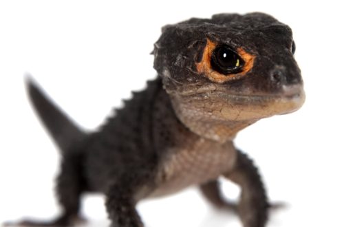
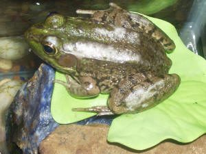
“Dropsy” of frogs, newts, axolotls and other amphibians
A lot of amphibian owners have experienced the fact that their pets began to develop “dropsy”, which is often called ascites. This is not very correct from the point of view of physiology, since amphibians do not have a division into the chest and abdominal cavities of the body due to the lack of a diaphragm, and ascites is still an accumulation of fluid in the abdominal cavity. Therefore, it is more correct to call the “dropsy” of amphibians a hydrocelom.
The edematous syndrome manifests itself in the form of a developing hydroceloma (accumulation of fluid sweating from the vessels in the body cavity) and / or generalized accumulation of fluid in the subcutaneous space.
Often this syndrome is associated with a bacterial infection and other processes that disrupt the protective function of the skin in maintaining homeostasis (the constancy of the internal environment of the body).
In addition, there are other causes of this syndrome, such as tumors, diseases of the liver, kidneys, metabolic diseases, malnutrition (hypoproteinemia), unsuitable water quality (for example, distilled water). With a lack of calcium in the body, the frequency and strength of heart contractions also decrease, which in turn leads to subcutaneous edema.
There are still many other yet unexplored causes of this syndrome. Some anurans sometimes experience spontaneous edema, which disappears spontaneously after a while. Some anurans also have subcutaneous edema, which may or may not have hydrocelom.
In addition, there are localized edemas, which are mainly associated with dysfunction of the lymphatic ducts due to trauma, injections, blockage with uric acid salts and oxalates, protozoan cysts, nematodes, compression due to an abscess or tumor. In this case, it is best to take edematous fluid for analysis and check for the presence of parasites, fungi, bacteria, salt crystals, cells that indicate inflammation or tumors.
If no signs of serious disease are found, then many amphibians live quietly with such localized edema, which may spontaneously disappear after some time.
Hydrocoelom is also found in tadpoles and is often associated with viral infections (ranaviruses).
To diagnose the causes of edema, sweating fluid and, if possible, blood are taken for analysis.
As a rule, for treatment, the veterinarian prescribes antibiotics and diuretics and, if necessary, drains excess fluid through punctures with a sterile needle.
Maintenance therapy includes saline baths (eg, 10–20% Ringer’s solution) to maintain electrolyte balance, which is very important for amphibians. It has been proven that the use of such salt baths along with antibiotics increases the percentage of recovery, compared with the use of antibiotics alone. Healthy amphibians maintain their own osmotic balance in the body. But in animals with skin lesions, bacterial diseases, kidney lesions, etc., the permeability of the skin is impaired. And since the osmotic pressure of water is usually lower than in the body, the permeability of water through the skin increases (water inflow increases, and the body does not have time to remove it).
Very often, edema is associated with severe lesions in the body, so treatment does not always have a favorable outcome. It must be remembered that it is better to consult a specialist at the very beginning of the disease.
At the same time, before going to the doctor, it is necessary to measure the temperature, pH and hardness of the water in which the pet is kept, since for some species this is a very important aspect.