
Breast tumors in dogs and cats

One of the most common reasons for visiting a veterinary oncologist is lumps in the abdomen in cats and dogs. As a rule, these are tumors of the mammary glands. The disease is more often recorded in elderly animals, older than 7 years. However, it also occurs in younger people. Unneutered bitches and cats are at greater risk of education. Males and cats are affected in rare cases, and in them the process is malignant. In bitches, about 40-50 percent of cases are benign, and in cats, 90% of cases are malignant – breast cancer. How to recognize the disease in time?
Contents
Symptoms of breast tumors
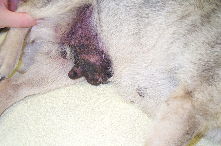
The onset of the disease may be imperceptible, especially in animals with thick hair. In the early stages, the owner can, when stroking the abdomen or probing the mammary glands, detect seals, they can be very small, the size of a pea. However, this is already cause for concern. The breasts may also be hot. There may be discharge from the nipples of a transparent, yellow or reddish hue. In later stages, the owner may find large growths that are often ulcerated, foul-smelling, and oozing. The general condition of the animal may be worsened: lethargy, lack or decrease in appetite, and so on. It is necessary to consult a doctor to clarify the diagnosis, since, for example, mastitis or a false pregnancy can occur with similar symptoms. The most dangerous symptoms that require prompt medical attention:
- rapid tumor growth
- change in the shape and color of education
- pain, redness, swelling
- the appearance of erosions and ulcers
Causes of the formation of tumors of the mammary glands
- As a rule, tumors are hormone-dependent. There are a lot of studies that castration before the first estrus (yes, castration is the removal of the uterus, ovaries, testes – regardless of gender) reduces the risk of developing breast tumors (BM) to 0,5%. If you castrate after the first heat – 8%, 26% after the second heat, after the third – castration does not affect in any way.
- In males, a severe hormonal imbalance can lead to pathology.
- The constant giving of drugs to the animal to suppress sexual desire also increases the chances of developing oncology.
- False puppies in bitches can also lead to changes in the mammary gland. Mastitis, mastopathy develop, which in the future can lead to cancer.
- Animal obesity.
Why are neoplasms dangerous?
The main danger of AMF is in metastasis. Altered cells spread through the blood or lymphatic vessels to the organs and tissues of the whole body, the lungs are most affected. Animals die from dysfunction of internal organs, exhaustion, lack of oxygen and other causes that arise due to oncology. Also, the opened tumors are the gates of infection, can fester and cause sepsis – blood poisoning.
Stages of development of breast tumors
Breast cancer staging is based on:
- state of the primary focus;
- the state of the tumor itself;
- the presence of altered lymph nodes;
- the presence of distant metastases.
It is believed that the criterion for the unfavorable behavior of the tumor is the size of the tumor: for cats it is 3 centimeters or more, for dogs of medium breeds 5-7 centimeters or more.
Stage 1 – a small seal or bump up to 1 cm in diameter, metastases are not detected. Stage 2 – neoplasm up to 3 cm in diameter, no signs of metastasis. Stage 3 – a larger formation up to 5 cm in diameter, may darken on the surface and in the deeper layers of the ulcer, which can bleed, there are metastases in the lymph nodes. Stage 4 – the tumor is larger than 5 cm in diameter. There are metastases in more distant areas of the body, more often in the lungs. Less commonly, veterinary oncologists encounter metastasis to the liver, spleen, pancreas, and bone tissue. It can be quite difficult to determine the stage of development by eye. To make a decision on the right treatment, a number of diagnostic procedures will be required.
Diagnostics
- Manual examination of the animal. Palpation of the mammary glands, external lymph nodes.
- Auscultation. Listening for murmurs in the lungs.
- Blood tests (biochemical and clinical). Assessment of the general functional state of the body.
- Ultrasound of the abdominal and thoracic cavity. Identification of structural changes in organs, the presence of large metastases.
- Chest x-ray at four! projections. Assessment of the state of the lung tissue, detection of metastases. One picture is not enough for a good diagnosis.
- Cytological examination allows you to make a preliminary diagnosis.
- Histological examination of the removed tumor will help to accurately determine the type of neoplasm, whether it is malignant or not.
- Cancer search using computed tomography. Alternative to x-ray and ultrasound, but performed under general anesthesia.
Treatment
Treatment depends on the stage of oncology, the general condition of the animal, concomitant diseases. At stages 1 and 2, surgeons often recommend a mastectomy – an operation to remove the mammary gland. More often, the entire ridge of glands is removed (unilateral mastectomy), sometimes (especially in the early stages) a partial mastectomy is performed, resection of only certain packets of glands. If the lesions are on both sides, then the operation is carried out in several stages, because the intervention is quite voluminous, painful and a supply of skin is required to tighten the edges of the wound. It is also recommended to castrate the animal at the same time. Often, surgeons detect changes in the tissues of the uterus and ovaries. In such a situation, the operation can take place in three stages. It is important that the oncologist who performs the operation understands ablastics – that is, he knows the rules for removing the tumor so as not to leave cells that can multiply again and so that metastasis does not occur. Resection of the neoplasm is carried out with a large seizure of surrounding tissues and with the removal of a nearby lymph node. After the operation, the animal is placed a special drainage tube in the area of the seam, into which the drug is injected for pain relief. Also, a cat or dog receives anti-inflammatory and analgesic drugs systemically. Chemotherapy is used in case of impossibility of surgical treatment or after determining a specific type of neoplasm, if necessary. There are many different protocols. The oncologist selects it individually, based on the characteristics of the patient. The life span of the appearance of breast tumors depends on the stage and extent of the spread of the process. Detection in the early stages allows starting effective treatment, which allows to completely remove the tumor and ensure a long-term remission – from 3-5 years or more. If the condition of the animal is so severe that none of the above methods is suitable, then the owners decide to carry out euthanasia or manipulation to improve the quality of life. Postoperative period Possible complications after surgery
- Suture infection
- Divergence of the sutures, most often occurs in the axillary and inguinal regions due to the large amount of tissue removed and the high mobility of the suture in these areas
- Tumor recurrence or spread of cancer that was not diagnosed before and during surgery
To prevent licking and infection of the sutures, a postoperative blanket and collar are put on, and restriction of mobility is also required for the healing time of the sutures, about 2 weeks. It is better to leave the animal for the first few days after the operation in the hospital for quality care and procedures. Most pets are discharged from the hospital 1-5 days after surgery, depending on the extent of the surgery and the condition of the patient. Most animals do not require any additional manipulations already 3-5 days after the operation. Patients are invited to a second appointment with an oncologist and a surgeon 12-16 days after the operation for a second examination and removal of sutures on the skin.
Prevention
The surest solution would be to castrate the pet before the onset of puberty, especially if the animal is not of breeding value. If the animal is not neutered, examine it more often, pay attention to the mammary glands of your cats and dogs, especially if they are already middle or old. Conduct a medical examination of your pet annually, this undoubtedly helps to identify and begin treatment not only for breast tumors, but also for other diseases earlier. Regular visits to the doctor with animals older than 6 years, timely diagnosis and treatment of tumors in the early stages reduce the risk of animal death from cancer.


You May Also Like

How to wean a dog from pulling on a leash: tips from Emily Larham

How to help a dog with a sensitive stomach?