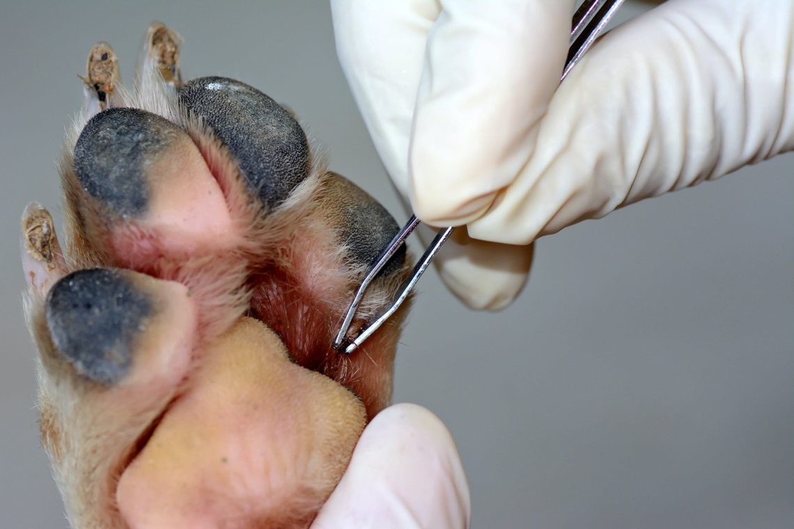
Babesiosis in dogs: symptoms
In recent years, there have been cases when babesiosis in dogs occurs without characteristic clinical signs and without a lethal outcome. However, when examining blood smears stained according to Romanovsky-Giemsa, babesia are found. This indicates the carriage of the pathogen. The diagnosis, as a rule, is made completely different: from poisoning to cirrhosis of the liver. Of particular interest is Babesia among stray city dogs. The presence of the freely circulating pathogen Babesia canis in a population of stray dogs is a rather serious link in the epizootic chain of the disease. It can be assumed that these animals are a reservoir of the parasite, contributing to its preservation. Thus, we can conclude that a stable parasite-host system has developed in the stray dog population. However, at this stage it is impossible to determine whether this happened due to the weakening of the pathogenic and virulent properties of Babesia canis or due to the increased resistance of the dog’s body to this pathogen. The incubation period for infection with a natural strain lasts 13-21 days, for experimental infection – from 2 to 7 days. In the hyperacute course of the disease, dogs die without showing clinical signs. The defeat of the body of the dog Babesia canis in the acute course of the disease causes fever, a sharp increase in body temperature to 41-42 ° C, which is maintained for 2-3 days, followed by a rapid fall to and below the norm (30-35 ° C). In young dogs, in which death occurs very quickly, there may be no fever at the onset of the disease. In dogs, there is a lack of appetite, depression, depression, a weak, thready pulse (up to 120-160 beats per minute), which later becomes arrhythmic. The heartbeat is amplified. Respiration is rapid (up to 36-48 per minute) and difficult, in young dogs often with a groan. Palpation of the left abdominal wall (behind the costal arch) reveals an enlarged spleen.

The mucous membranes of the oral cavity and conjunctiva are anemic, icteric. Intensive destruction of red blood cells is accompanied by nephritis. The gait becomes difficult, hemoglobinuria appears. The disease lasts from 2 to 5 days, less often 10-11 days, often fatal (N.A. Kazakov, 1982). In the vast majority of cases, hemolytic anemia is observed due to the massive destruction of red blood cells, hemoglobinuria (with urine becoming reddish or coffee-colored), bilirubinemia, jaundice, intoxication, damage to the central nervous system. Sometimes there is a lesion of the skin such as urticaria, hemorrhagic spots. Muscle and joint pains are often observed. Hepatomegaly and splenomegaly are often observed. Agglutination of erythrocytes in the capillaries of the brain can be observed. In the absence of timely assistance, animals, as a rule, die on the 3rd-5th day of the disease. A chronic course is often observed in dogs that have previously had babesiosis, as well as in animals with increased body resistance. This form of the disease is characterized by the development of anemia, muscle weakness and exhaustion. In sick animals, there is also an increase in temperature to 40-41 ° C in the first days of the disease. Further, the temperature drops to normal (on average, 38-39 ° C). Animals are lethargic, appetite is reduced. Often there are diarrhea with bright yellow staining of fecal matter. The duration of the disease is 3-8 weeks. The disease usually ends with a gradual recovery. (ON THE. Kazakov, 1982 A.I. Yatusevich, V.T. Zablotsky, 1995). Quite often in the scientific literature one can find information about parasites: babesiosis, anaplasmosis, rickettsiosis, leptospirosis, etc. (A.I. Yatusevich et al., 2006 N.V. Molotova, 2007 and others). According to P. Seneviratna (1965), out of 132 dogs examined by him for secondary infections and infestations, 28 dogs had a parasitic disease caused by Ancylostoma caninum 8 – filariasis 6 – leptospirosis 15 dogs had other infections and infestations. The dead dogs were exhausted. Mucous membranes, subcutaneous tissue and serous membranes are icteric. On the intestinal mucosa, there are sometimes point or banded hemorrhages. The spleen is enlarged, the pulp is softened, from bright red to dark cherry color, the surface is bumpy. The liver is enlarged, light cherry, less often brown, the parenchyma is compacted. The gallbladder is full of orange bile. The kidneys are enlarged, edematous, hyperemic, the capsule is easily removed, the cortical layer is dark red, the brain is red. The bladder is filled with urine of red or coffee color, on the mucous membrane there are pinpoint or striped hemorrhages. The cardiac muscle is dark red, with banded hemorrhages under the epi- and endocardium. The cavities of the heart contain “varnished” non-clotting blood. In the case of a hyperacute course, the following changes are found in dead animals. The mucous membranes have a slight lemon yellowness. The blood in large vessels is thick, dark red. In many organs, there are clear pinpoint hemorrhages: in the thymus, pancreas, under the epicardium, in the cortical layer of the kidneys, under the pleura, in the lymph nodes, along the tops of the stomach folds. The external and internal lymph nodes are swollen, moist, gray, with noticeable follicles in the cortical zone. The spleen has a dense pulp, giving a moderate scraping. The myocardium is pale gray, flabby. The kidneys also have a flabby texture. The capsule is easy to remove. In the liver, signs of protein dystrophy are found. The lungs have an intense red color, a dense texture, and thick red foam is often found in the trachea. In the brain, smoothness of the convolutions is noted. In the duodenum and anterior part of the lean mucous membrane reddened, loose. In other parts of the intestine, the surface of the mucosa is covered with a moderate amount of thick gray mucus. Solitary follicles and Peyer’s patches are large, clear, densely located in the thickness of the intestine. As a feature, it should be noted the absence of hemosiderosis – an indicator of increased breakdown of erythrocytes (V.L.
See also:
What is babesiosis and where do ixodid ticks live
When can a dog get babesiosis?
Babesiosis in dogs: diagnosis
Babesiosis in dogs: treatment
Babesiosis in dogs: prevention


