Third eyelid in cats: causes of pathologies and treatment
Contents
third eyelid in cats
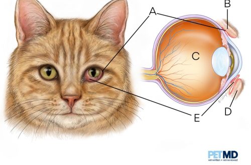
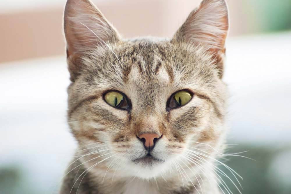
Usually, the owner immediately notices if a whitish film suddenly appears in front of the cat’s eyes. In fact, this eyelid, or, as it is also called, the nictitating membrane, is present in all cats. It is just normal that it is neatly folded in the inner corner of the eye and looks like a small white or colored crescent.
The functions of the third century are very important:
It houses one of the lacrimal glands, which helps to moisturize the surface of the eye.
On its inner surface are the structures of the immune system – lymphoid tissues.
It distributes the tear over the surface of the eye and helps remove debris, mucus, and pathogens.
Protects the surface of the eye from damage.
Pathologies of the third eyelid in cats
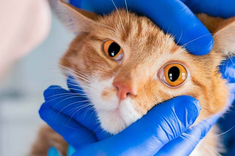
It is by no means always that changes in the third eyelid will be associated with its damage, inflammation or eye disease. In cats, they can also signal a systemic problem with the body, an infectious or neurological disease.
Third eyelid prolapse in a cat
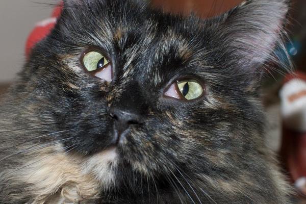
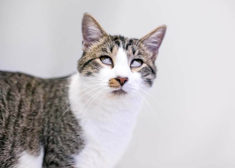
In another way, it is called prolapse or protrusion. The prolapse causes the cat’s third eyelid to become prominent and cover part of the eye. Such prolapse in a cat can be unilateral and bilateral, that is, affect one or both eyes.
Bilateral fallout can be seen:
Normal during sleep, as well as after exposure to anesthesia, while the animal has not yet fully regained consciousness.
For eye diseases: conjunctivitis, corneal erosion and ulcer, keratitis, uveitis, glaucoma, etc.
With significant dehydration, fever, exhaustion and other ailments.
Idiopathic prolapse, the cause of which is not known for certain.
Unilateral fallout may be accompanied by:
Eye disease (ulcer, keratitis, neoplasia, glaucoma, uveitis), if the pathological process has affected only one eye.
Horner’s syndrome. It accompanies various neurological pathologies: otitis media, damage to the brain and spinal cord, etc. In some cases, the manifestation may be bilateral.
Injury
The third eyelid can be injured like any other structure in the eye. The most common cause of injury will be cat’s claws. This happens during a fight or game with another animal, and also accidentally when a cat scratches and rubs its eyes, head, ears.
Outwardly, it will often look like inflammation. In this case, the cat’s third eyelid swells and turns red. With an injury, a rupture or partial detachment can occur, and sometimes damage to its cartilage.
Inflammation of the third eyelid in cats
Inflammation of the third eyelid in a cat will most often be observed with conjunctivitis – inflammation of the conjunctiva. This is a thin and delicate mucous membrane that covers the inside of the eyelids, the eyeball to the cornea and directly the third eyelid.
With inflammation, the third eyelid thickens, swells, turns red. Lymphoid tissues – follicles – on its inner surface can also become more pronounced and look like a scattering of bubbles.
The most common causes of conjunctivitis in cats are viruses and bacteria: feline herpes, calicivirus, chlamydia, and mycoplasmas. More rare: irritating external factors (dust, smoke), foreign bodies, allergies.
neoplasm
They can directly affect the third eyelid, but are quite rare. Outwardly, you can observe a change in its size, shape and color. This disease is found mainly in older animals. Most often, the defeat will be unilateral.
Adenoma of the third eyelid in a cat
This is an inflammation and benign enlargement of the gland of the third eyelid in a cat. Often it will be accompanied by its simultaneous loss – prolapse. Outwardly, the adenoma of the gland looks like a pink or red tubercle in the corner of the eye. In cats, this problem is less common than in dogs, and the so-called brachycephalic breeds with a shortened, flattened muzzle are more susceptible to it: Persian, exotic, British, Scottish.

Hall
This pathology of the third eyelid occurs, as a rule, in dogs of large breeds. In cats, this is a fairly rare problem. The shape of the third eyelid helps to maintain thin cartilage, sometimes it curves. Outwardly, this pathology will be similar to prolapse or trauma of the third eyelid.
Symptoms
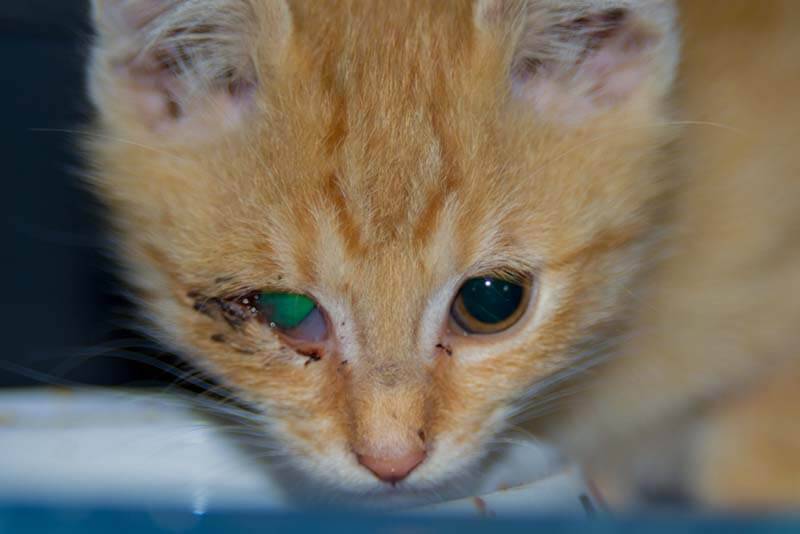
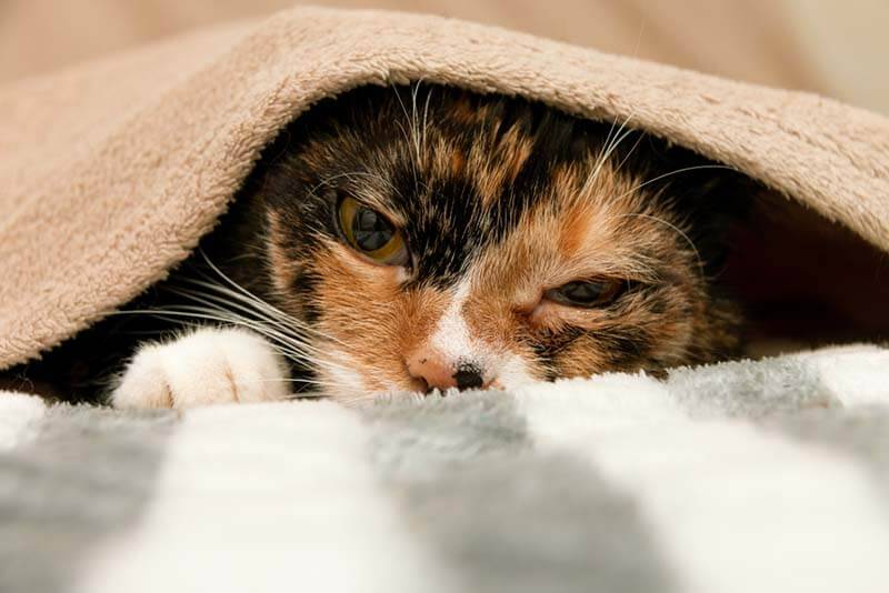
With eye diseases, in addition to prolapse of the third eyelid, cats often experience redness and swelling of the conjunctiva, photophobia and blepharospasm – when the animal squints its eyes or does not open them at all. Often there is severe lacrimation, mucous or purulent discharge from the eyes.
With trauma and neoplasm of the nictitating membrane, there may be bleeding, squinting of the eye, increased lacrimation.
The third eyelid itself becomes swollen, inflamed, with a torn or uneven edge.
Adenoma of the gland of the third eyelid often looks like a small pink or red ball in the inner corner of the eye. A similar picture will be with a prolapse of the gland and with a fracture of the cartilage of the third century.
Horner’s syndrome is accompanied by drooping eyelids, retraction of the eyeball, a change in the size of the pupil, and sometimes other neurological symptoms: a forced tilt of the head to one side, impaired coordination.
Diagnostics
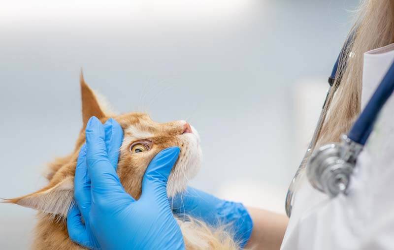
Since there are many reasons for problems with the third eyelid, it is possible to draw up an approximate diagnostic plan only upon examination by a doctor.
If, in addition to prolapse of the third eyelid, the pet has other symptoms of general malaise: vomiting, fever, dehydration, it will be important to find out the original cause of the disease. This may require an ultrasound examination of the abdominal cavity, blood tests.
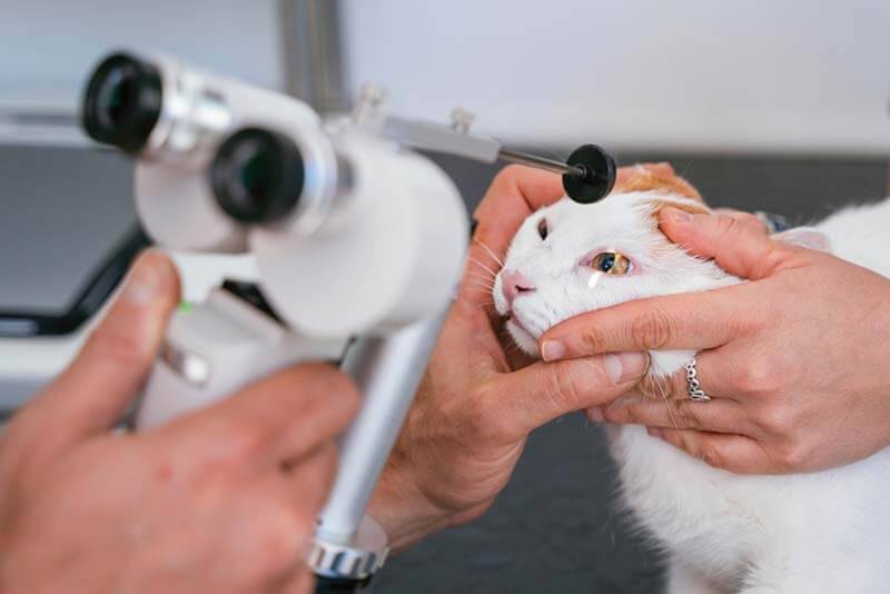
With an adenoma of the gland of the third century, as well as its injury, often only an examination by an ophthalmologist is necessary. Often, the examination is carried out under general anesthesia, but, if the temperament of the animal and the volume of the pathology allows, under local anesthesia with the help of special eye drops.
If the prolapse of the third eyelid is accompanied by symptoms such as a change in the size of the pupils, drooping of the eyelids, impaired coordination, then an examination by a neurologist will be required.
With increased lacrimation, squinting of the eyes, redness and swelling of the conjunctiva, mucopurulent discharge, it will be necessary to exclude eye diseases: conjunctivitis, keratitis, corneal erosion, etc. This will require a test with fluorescein – a special solution that glows in ultraviolet rays and helps to identify damage cornea, as well as to measure the production of tears, intraocular pressure.
Sometimes specific studies are needed for specific pathogens: feline herpes virus, chlamydia, mycoplasma.
If a neoplasm of the third eyelid is suspected, cytology or histology may be required, when tissue cells or specially prepared tissue sections are examined under a microscope.
Why are these diseases dangerous?
Regardless of what pathology caused the changes in the nictitating membrane in a cat, it must be identified, and without delay. A seemingly insignificant eye problem with untimely or incorrect treatment can result in serious complications and lead to partial or complete loss of vision.
Any eye disease causes discomfort and pain, as the cornea is very sensitive.
Adenoma of the gland of the third eyelid, even if it prolapses, does not pose a direct threat to the life of the pet, but such a gland cannot function correctly. Plus, the animal perceives it as a hindrance and tries to rub and scratch the eye, risking injuring it and significantly aggravating the problem.
An injury to the third eyelid can lead to partial or complete loss of its functionality.
The neoplasm is much easier to remove when it is still small and has not affected other structures of the eye.
If Horner’s syndrome became the cause of the protrusion of the nictitating membrane, then this indicates the presence of a serious neurological disease in the animal.
How and how to treat pathologies of the third eyelid in cats
When the third eyelid falls out against the background of other pathologies, the primary problem, for example, an infectious disease, otitis media, etc., is subject to treatment.
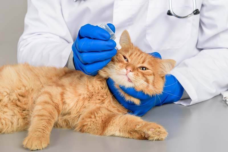
If inflammation of the third eyelid is associated with conjunctivitis, antibacterial and moisturizing eye drops are used. Sometimes it is necessary to use an antibiotic systemically – inside.
With conjunctivitis caused by feline herpes, antiviral therapy is additionally prescribed – preparations based on famciclovir.
Other eye conditions that show the cat’s third eyelid may require more specific treatment. For example, with significant lesions of the cornea – erosion, ulcers – a procedure such as tarsorrhaphy can be used, when the third eyelid is temporarily sutured to the upper eyelid. This is necessary to protect the eye and its speedy healing.

In case of adenoma and prolapse of the gland of the third eyelid, the doctor repositions it and, if necessary, sutures it to keep it in the correct position. Also, surgical treatment will be required for a crease.
With a minor injury without separation, healing can occur against the background of the use of local drops: moisturizing, analgesic and antibacterial.
In severe cases of injury, surgical repair of the integrity of the third eyelid may be required. They try to preserve it as much as possible, since when the third eyelid is removed, the animal will begin the gradual development of dry keratoconjunctivitis – inflammation associated with chronic insufficient hydration of the eye.
Removal of the third eyelid is necessary for an extensive neoplasm, especially if a malignant process is suspected.
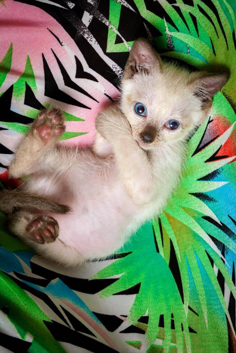
Third eyelid in kittens
In kittens, the reasons for the change in the third eyelid are the same as those discussed above, with the exception, perhaps, of neoplasms.
Somewhat more often, compared with adult pets, they can be diagnosed with eye injuries, including those of the third eyelid.
This is due to games between littermates and active knowledge of the surrounding world.
Kittens are more susceptible to infections, including feline herpes virus.
In babies, herpes is more severe and, in advanced cases, can lead to the loss of one or both eyes.
Also, kittens and young animals have a higher risk of stricture (narrowing) of the third eyelid – its growth to the conjunctiva and cornea against the background of inflammation. For this reason, it is important to start treatment of all eye diseases in kittens in a timely manner and carry out carefully.
Prevention
By and large, the prevention of problems with the third eyelid comes down to the general prevention of safety and maintaining the health of the pet:
Comfortable and safe conditions of detention
Trimming the nails of cats when they are kept together and avoiding regular conflicts
Timely vaccination against viral diseases
Prevention of free range on the street
Promptly contact your veterinarian if you are symptomatic.
Home
All members of the cat family have a third eyelid, but normally it is almost invisible.
It is an important structure of the eye that performs protective, moisturizing and immune functions.
A problem with the third eyelid can be both a symptom and an independent disease.
An independent disease of the third century can be attributed to: adenoma and prolapse of the gland, trauma, neoplasm.
In pathology, the third eyelid becomes noticeable, covers part of the eye, turns red, swells, changes shape (for example, with gland adenoma, neoplasm, trauma). In addition, there may be discharge from the eyes.
Treatment is based on the diagnosis. In case of injury and inflammation, antibiotic therapy, moisturizers are needed. Adenoma of the gland of the third eyelid and its prolapse require reduction and fixation in the correct position. Removal of the third eyelid is carried out only in extreme cases – with serious injuries with the impossibility of recovery or neoplasms.
Answers to frequently asked questions
Sources:
Oleinik V.V. Veterinary ophthalmology – atlas, 2013
R. Rees. Small Animal Ophthalmology, 2006
H. Featherstone, E. Holt. Ophthalmology of dogs and cats, 2018
Vasilyeva E.V. Pathologies of the third century // Zooinform 2019
16 May 2022
Updated: 16 May 2022


