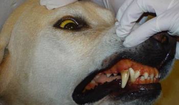
Demodicosis in dogs
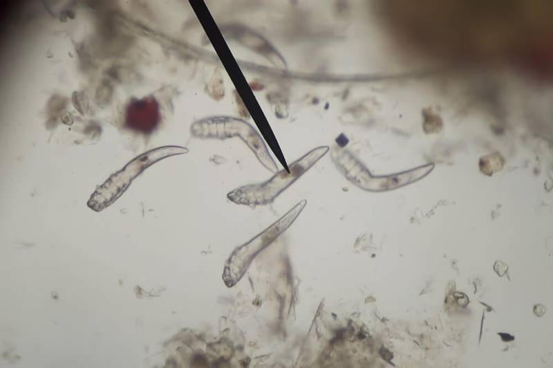
Demodex mite — it is a normal inhabitant of the skin in dogs and can be found in the skin and ear canals even in healthy animals. It gets on the skin of newborn puppies from the mother in the first 2-3 days of life. It is impossible to get infected with demodicosis from a sick dog; intrauterine transmission is also excluded. In the study of tissues of dogs that died due to various diseases, these parasites were also found in the internal organs, in urine, feces and blood. But such findings are considered to be accidental, since the tick breathes oxygen and, accordingly, could not live inside the body. The drift of ticks into the internal organs occurs with blood and lymph from the focus of inflammation. Outside the body, these mites also cannot live.
Almost 80% of cases of demodicosis are observed in purebred dogs, only 20% occur in outbred animals. There is also a breed predisposition: for example, Scottish Terrier, Shar Pei, Afghan Hound, Great Dane, English Bulldog, West Highland White Terrier, Doberman get sick more often than others.
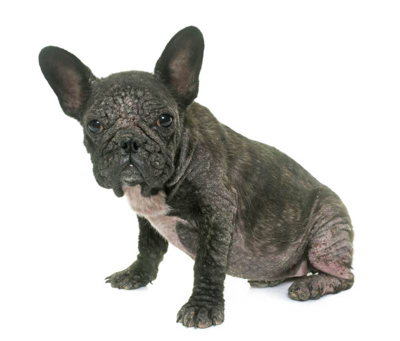
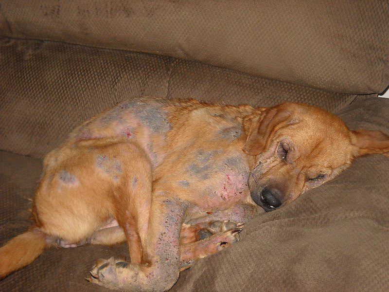
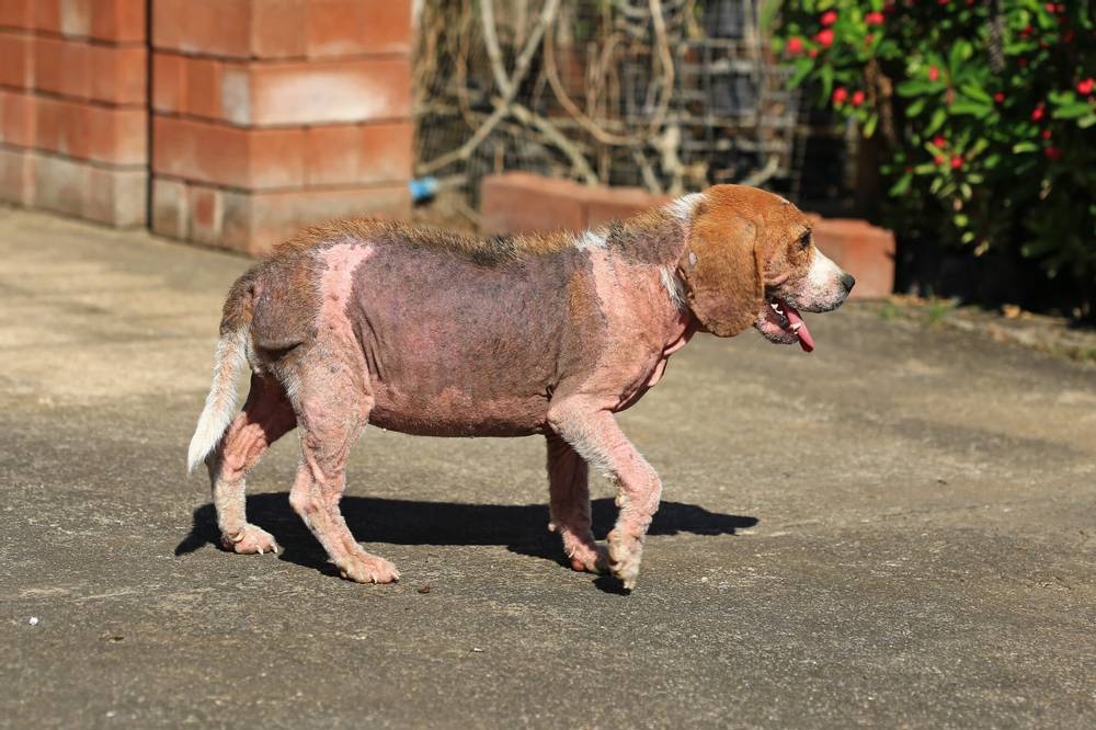
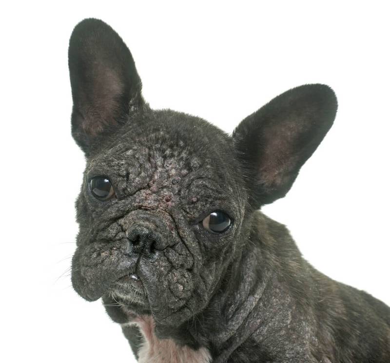
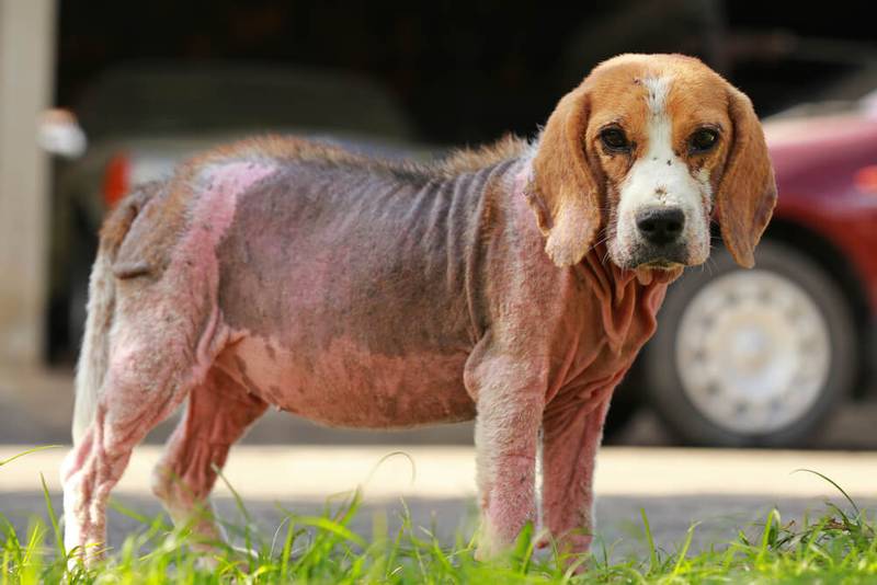
Photo of demodicosis in dogs
Contents
Causes
The main reason for the development of demodicosis in dogs — It’s reduced immunity. Immunity can be reduced against the background of various diseases present in the animal: infectious, inflammatory, diabetes mellitus, malignant tumors, endocrine disorders, as well as during estrus and pregnancy in bitches. The use of various drugs that have an immunosuppressive effect (for example, drugs from the group of glucocorticosteroids) also leads to a decrease in immunity. Poor conditions for keeping a dog, poor-quality feeding, lack of exercise, crowded content, lack of warm rooms for keeping in the cold season — all this contributes to a decrease in the body’s own immune forces and can become a factor in the development of demodicosis. Another cause of demodicosis — a genetic defect, that is, inherited. This defect affects lymphocytes (cells of the immune system), which leads to the uncontrolled reproduction of parasites.
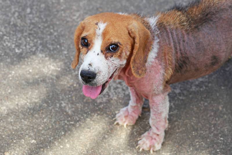
Symptoms of demodicosis in dogs
The first sign to suspect the development of the disease in your dog is — this is the appearance of alopecia, that is, areas of the body with hair loss and a violation of the growth process of new ones. Other symptoms of demodicosis in a dog can be redness and peeling of the skin, the formation of pustules. Particular attention should be paid to the skin around the eyes, lips. In the initial stage of demodicosis, the dog will not itch, and these lesions will not cause concern to the animal. Itching appears only when a secondary bacterial or fungal infection is attached to existing lesions. Staphylococcus bacteria (mainly Staphylococcus pseudintermedius) can be found most often, streptococci, rod-shaped bacteria and yeast fungi (genus Malassezia) are somewhat less common. In especially neglected cases, there may be depression of general well-being, refusal to eat, the animal may even die from sepsis.
Types of demodicosis
According to the prevalence of lesions, one can distinguish between localized (a small number of lesions on the body) and generalized demodicosis (capturing large surfaces of the skin). By age, it is divided into juvenile (demodicosis in puppies) and adult dogs. By type of clinical manifestation — pustular (pyodemodecosis), papular (nodular), squamous (scaly) and mixed.
Localized
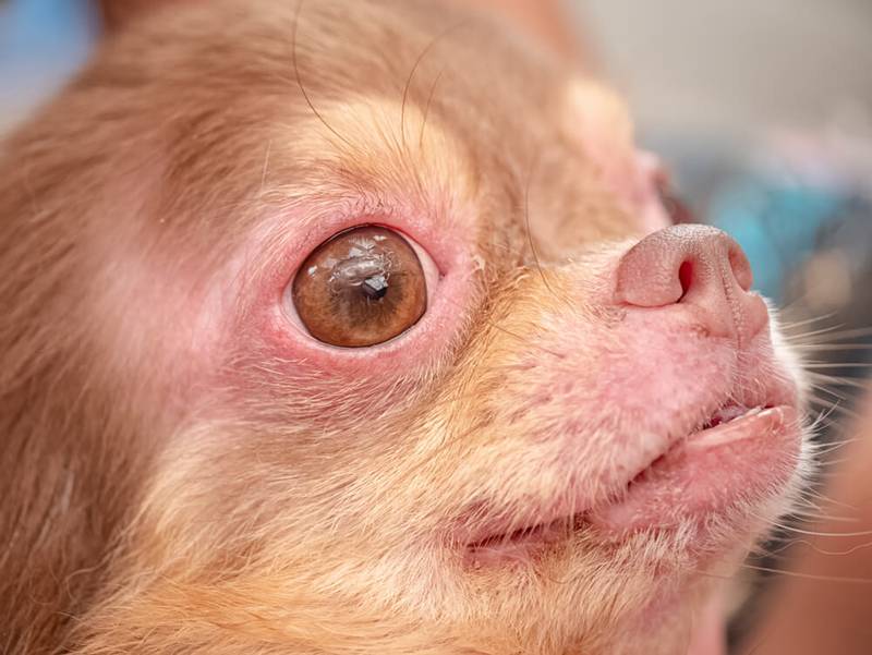
Most often it can be found in young dogs (up to about 1 year old). According to modern data, demodicosis is considered localized if there are five or less lesions on the body with a diameter of up to 2,5 centimeters. These lesions are well-demarcated areas, without hair, with or without redness, and peeling is also possible. The skin may have a bluish-gray tint, comedones (black dots) and an unpleasant odor are sometimes noted. Most often, such lesions are found on the muzzle, head, neck, front legs. You can find the characteristic “demodectic” glasses in the form of redness around the eyes. About 10% of cases of a localized course turn into a generalized form.
Generalized
The clinical picture is similar to localized demodicosis, but it captures more areas of the dog’s skin. It is customary to call generalized demodicosis if there are more than 5 lesions, or these lesions are more than 2,5 centimeters, or if one part of the body is affected as a whole (the entire muzzle, the entire leg, etc.). Clinical symptoms include baldness, peeling, comedones, darkening of the skin. Most likely, the addition of a secondary bacterial or fungal flora, which causes the appearance of pimples and pustules, boils (inflammation in the area of the hair root, that is, already in the deeper layers of the skin) and fistulas. With this variant of the course, itching will be an integral part of the disease, and over time it will develop into a truly painful sensation. In extremely advanced cases, one should expect an increase in lymph nodes, a decrease in appetite, and depression of the general condition. Without treatment, the animal will die fairly quickly.
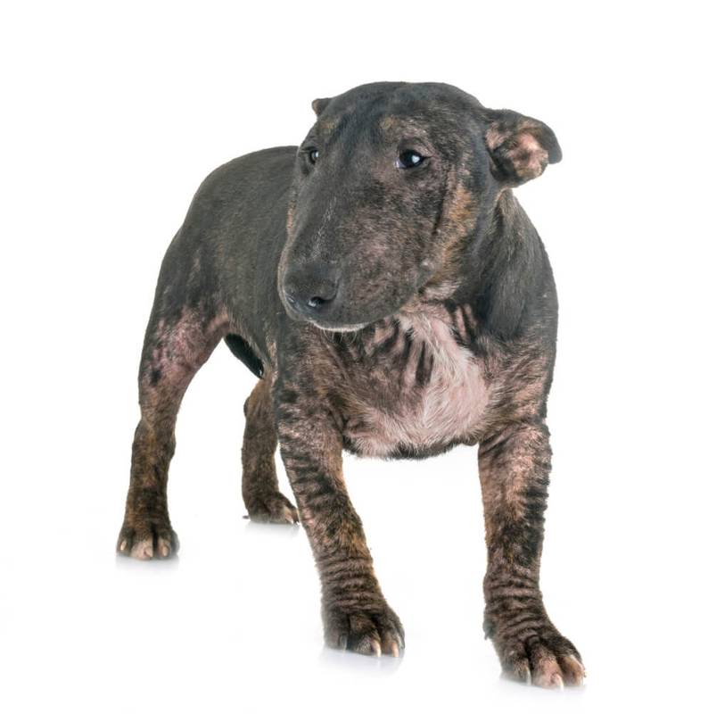
Generalized demodicosis also includes mite damage to the limbs of a dog. — pododemodecosis. You can observe swelling of the paws, darkening of the skin, interdigital cysts, fistulous passages with outflows of a different nature from them, lameness due to pain. The dog will constantly lick the limbs, especially the pads and between the toes. May become aggressive when trying to wash their paws after a walk. Podomodedecosis is difficult to treat.
In rare cases, even the ear canals are affected, causing otitis externa (otodemodicosis). This type of lesion also refers to the generalized form. You can observe redness of the inner surface of the ears, brown discharge, an unpleasant smell from the ears. At the same time, the dog can shake its head, rub its ears against various objects, and also scratch the ears and the area next to the ears (cheeks, neck).
Juvenile
Juvenile demodicosis is a disease of puppies aged mainly from 6 to 12 months. This type of demodicosis is almost always caused by a hereditary defect in the immune system, that is, one of the parents was also sick. The organism of these puppies is not able to independently regulate the number of ticks, as a result of which their population increases and they cause clinical manifestations of the disease. Such animals must be removed from breeding to prevent the spread of the disease. The rest of the clinical signs will depend on the form of the course of the disease (localized or generalized).
adult animals
In adult animals, the development of the disease will often be associated with a decrease in immunity against the background of the underlying disease. Therefore, when demodicosis is detected in adult dogs, a thorough examination of general health is also necessary: a complete physical examination and additional studies. Particular attention should be paid to the search for diseases such as diabetes mellitus, hypothyroidism, hyperadrenocorticism, and malignant tumors. According to the data, successful treatment of the underlying disease gives a good remission to demodicosis. However, more than half of the dogs that underwent a complete examination showed no other diseases. Another cause of demodicosis in adult animals is the long-term use of immunosuppressive drugs that were prescribed to treat the primary disease.
pustular
This form is characterized by the appearance of pustules on the skin. These pustules burst after a while, their contents flow out and dry out. The skin may turn red or darken, it becomes wrinkled and firmer, and an unpleasant odor appears. Infection of the skin occurs quickly enough and spreads to other parts of the body that were not originally affected by the parasite.
Papular
With this form, rounded, most often red and distinctly limited nodules can be observed in various parts of the body, their diameter can reach 1-6 millimeters. These nodules may be itchy in the dog, but they may also not cause concern at all.
Squamous
With the squamous type, small, mosaic lesions appear on the skin of the dog, covered with bran-like scales. Over time, they begin to merge, in these places there is increased hair loss.
Mixed
This type of lesions includes all of the above clinical signs (papules, pustules and scales) and can be quite severe, depressing the general well-being of the animal.
Diagnostics
The diagnosis is made comprehensively, taking into account the history (complaints according to the owner, medical history), physical examination, and laboratory tests. The main method of confirming the diagnosis is microscopy of skin scrapings. Scraping is necessary from all affected areas of the body. The scraping should be deep enough, carried out with a scalpel until the first drops of blood appear, since the tick sits in the deep layers of the skin (hair follicle). Trichoscopy (examination of plucked hairs) or an adhesive test (taking material for examination using a narrow tape of adhesive tape) may also be useful. If there are whole pustules on the body, it is imperative to conduct a microscopy of their contents. To make a diagnosis, you need to find a large number of ticks at different stages of their development. The discovery of just one tick can be an accidental finding, but still should not be completely ignored. In such cases, scrapings are recommended to be repeated after a while (2-3 weeks) to clarify the diagnosis. If otodemodecosis is suspected, then microscopy of the contents of the external auditory canals is performed. In particularly doubtful cases, a skin biopsy with its histological examination may be suggested. Also, in doubtful cases, a trial treatment may be offered by the doctor, even if the diagnosis could not be confirmed at the initial appointment.
Treatment of demodicosis in dogs
In modern regimens for the treatment of demodicosis in dogs, the safest oral preparations from the isoxazoline group (fluralaner, afoxolaner, sarolaner) are used. Such drugs are also used to prevent flea and tick bites on an ongoing basis, without the risk of harming the body when used according to the instructions. The scheme of treatment with them can be different and depends on the degree of damage to the dog with demodicosis and the specific drug chosen.
In the absence of financial or other opportunities to use such drugs, a classic treatment regimen using drugs of the avermectin group can be applied. These injectables work well when taken orally, but have more side effects (drooling, lethargy, staggering gait, convulsions, and coma). Their use is contraindicated in puppies under three months of age. There is also breed intolerance to drugs from this group in some dogs (collie, English shepherd dog, Australian shepherd, Scottish shepherd dog and their crosses). This is due to the presence of a defective gene in their body, due to which the drug molecule “remains” in the brain and cannot leave it, causing a wide range of neurological problems.
For the treatment of demodicosis, drugs from the amitraz group in the form of an aqueous solution can be used as baths on the entire surface of the body, but its use is also associated with possible side effects (lethargy, itching, urticaria, vomiting, refusal to eat, unsteady gait usually disappear after 12 -24 hours).
There is also evidence of the high effectiveness of macrocyclic lactones in the treatment of demodicosis, but this issue is still controversial. In the presence of a secondary infection, local preparations (various antibacterial ointments and shampoos) can be prescribed, in especially advanced cases, systemic antibiotics are prescribed in dermatological doses.
It is necessary to continue treating demodicosis in a dog until two consecutive negative scrapings are obtained with an interval of one month between them. Treatment may be extended for another month thereafter as a relapse prevention measure. Relapses in the generalized form of the course are not rare. Their treatment can be quite long, up to six months or longer. Such animals can even be euthanized.

Danger to humans
Demodex is a strictly specific parasite, that is, a species that parasitizes on dogs, but cannot parasitize on humans. And, as noted above, demodex is a normal inhabitant of the skin of an animal. It multiplies, causing disease, only in the conditions of a particular organism (due to a decrease in immunity or a genetic defect) and, accordingly, is not contagious.
Prevention
The best prevention of the occurrence of demodicosis is to maintain the dog’s immunity at a high level. This can be achieved by creating comfortable living conditions for her: quality food, regular exercise, care and affection. It is also necessary to conduct regular preventive examinations at the veterinarian to identify possible pathologies, especially for animals older than 7 years. All animals with a generalized form of demodicosis should not be bred, since with a high degree of probability the defective “demodectic” gene will be passed on to offspring. Such dogs can be castrated, which also prevents the occurrence of disease in bitches during estrus.
Possible complications
Complications with a localized form of the course of demodicosis and timely treatment, as a rule, are not observed. The main possible complications include secondary infection with bacterial and fungal agents. With untimely treatment, there will also be an increase in palpable lymph nodes, an increase in body temperature, general depression, refusal to eat, unbearable itching. This is followed by sepsis and death of the animal.
The article is not a call to action!
For a more detailed study of the problem, we recommend contacting a specialist.
Ask the vet
2 September 2020
Updated: February 13, 2021


