
Digestive diseases in guinea pigs
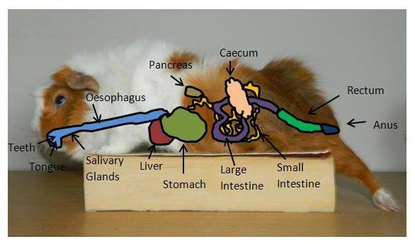
The digestive system of the guinea pig is very susceptible to disorders due to the large length of the intestine and the long passage of food through the intestines. Accordingly, guinea pig owners often bring guinea pigs to veterinarians with digestive disorders. The intestinal flora is sensitive to changes in feed composition. Replacing the usual food with a new one is recommended to be done very slowly if you bought a pig in a store or nursery. It is necessary to find out how the pig was fed before in order to avoid problems associated with a sudden change in diet.
Contents
- Enteritis
- E. coli
- salmonellosis
- Constipation
- Endoparasites
- Trichomoniasis
- Amoebiasis
- Coccidiosis
- Toxoplasmosis
- Fascioliasis
- Tapeworm (tapeworm) infection
- Enterobiasis (pinworm infection)
- Enteritis
- E. coli
- salmonellosis
- Constipation
- Endoparasites
- Trichomoniasis
- Amoebiasis
- Coccidiosis
- Toxoplasmosis
- Fascioliasis
- Tapeworm (tapeworm) infection
- Enterobiasis (pinworm infection)
- Viral infection of the salivary gland in guinea pigs
- Dental anomalies in guinea pigs
- Tympania in guinea pigs
Enteritis
The sensitive digestive system of the guinea pig is often affected by enteritis. The reasons for the violation of the composition of microorganisms in the intestine can be different. A severe disturbance of the intestinal flora is caused by a change in the composition of the feed, the lack of a sufficient amount of coarse fiber, oral antibiotics, or refusal to eat for many days.
The clinical symptoms are diarrhoea, bloating, and a loud bowel noise. When examining urine, the analysis of which is taken by squeezing the bladder, ketone bodies are found. Therapy consists in restoring a normally functioning intestinal flora. Therefore, within 36 hours after the onset of symptoms, only hay can be given as dietary food to animals. Of course, it must be of impeccable quality, since moldy food can also lead to enteritis. It is impossible to administer antibiotics orally, as this will disrupt the restoration of the intact intestinal flora. It is recommended to give guinea pigs intestinal bacteria. To do this, you need to dissolve the droppings of healthy guinea pigs in a small amount of water and inject this solution using a disposable syringe. Fluid loss due to diarrhea can be replaced by subcutaneous injection of glucose and electrolyte solutions. To restore the intact intestinal flora, the animal must necessarily take food, even artificially in case of refusal (see the chapter “Special Instructions”).
E. coli
Another type of infectious enteritis is caused by Escherichia coli. Changes in the intestinal flora can lead to a strong accumulation of Escherichia coli microorganisms, which are not usually found in the intestines of a guinea pig. The disease progresses rapidly, the animals develop bloody diarrhea and die within a few days.
salmonellosis
A special form of enteritis is salmonellosis. This disease can be latent, acute and chronic. Guinea pigs become infected with salmonellosis most often from the droppings of wild rabbits or mice, as well as through food. In an acute course, the disease is accompanied by severe diarrhea and leads to death within 24-28 hours; in the chronic nature of the disease, diarrhea is constantly repeated and there is no appetite. After the resistance test, antibiotics are administered parenterally to the animal. With the acute nature of the disease, the animal has no chance of recovery. Due to the risk of infection to humans, after any handling of guinea pigs with Salmonellosis, hands must be thoroughly washed and disinfected. Other pets and children should also not be allowed near them.
Constipation
Occasionally, guinea pigs are brought to the veterinarians who have not had a bowel movement in several days and are showing symptoms of severe abdominal pain; animals are very lethargic. The balls of litter accumulated in the intestines are well palpable. Treatment must be carried out very carefully in order to damage the very sensitive intestinal mucosa as little as possible. Therefore, strong laxatives should not be used. Using a disposable syringe, 2 ml of paraffin oil is administered orally to the animal, 1/4 tube of Mikroklist is injected into the rectum. 0,2 ml of Bascopan, injected under the skin, can support treatment. Gentle massage of the abdomen can stimulate intestinal motility and relieve pain.
If the above treatment does not work within a few hours, then an x-ray (possibly with barium sulfate) should be taken. In guinea pigs, closure of the intestinal lumen caused by various reasons was observed, in which surgical intervention was necessary. True, the chances of success here are limited.
Endoparasites
Diseases caused by endoparasites are very rare in guinea pigs, with the possible exception of coccidiosis, although they are widely described in the literature. In this case, we are often talking about autopsy data.
Trichomoniasis
Symptoms of trichomoniasis are diarrhea and weight loss. This disease is most often caused by Trichomonas caviae and Trichomonas microti. With a strong lesion, Trichomonas can cause inflammation of the intestines. They are easy to see in a smear of litter under a microscope. Treatment is with metronidazole (50 mg/1 kg body weight). The medicine must be mixed into the water, and it is better to feed the animals only with dry food, while making sure that the animals drink enough water.
Amoebiasis
The same treatment is done for amoebiasis caused by Endamoeba caviae or Endamoeba muris. Infection with amoebiasis occurs as a result of ingestion of cysts. The cyst can be detected by flotation. Amoebas also cause inflammation of the intestines, the manifestations of which are diarrhea and weight loss.
Coccidiosis
Coccidiosis is the most common disease in guinea pigs caused by endoparasites of the meria species group, Eimeria caviae. The first symptom is incessant diarrhea, and the droppings are often mixed with blood. Oocytes can be seen under a microscope: with a strong lesion – in a native preparation, with a weak one – using the flotation method. In this case, it is also better to mix the medicine into water. Animals should be fed exclusively with dry food, and a sufficient amount of liquid was ingested in the form of water. Sulfamethacin (7 g / 1 l of water) or (also within 1 days) 7% sulfamidine should be added to the water for 2 days.
Toxoplasmosis
The causative agent of toxoplasmosis, Toxoplasma gondii, has also been found in guinea pigs. However, an animal infected with toxoplasmosis cannot shed infectious oocysts. Since we no longer eat guinea pigs, human infection is ruled out.
Fascioliasis
Among flukes, only Fasciola hepatica is dangerous to guinea pigs. A guinea pig can become infected with them through grass or ants from an infected meadow. Veterinarians make such a diagnosis only in exceptional cases. Basically, this is the data of the autopsy. In the presence of such autopsy results, the owner should find another source of food for his animals in order to avoid infection with Fasciola hepatica in the future. Symptoms of fascioliasis are apathy and weight loss. However, they appear only in the case of a severe lesion, in which the treatment does not promise much success. With fasciolosis, pracicantel is prescribed (5 mg / 1 kg of body weight).
Tapeworm (tapeworm) infection
Tapeworms are extremely rare in guinea pigs. The most common are Hymenolepis fraterna, Hymenolepsis papa, and Echinococcus granulosus. As a medicine, give once (5 mg / 1 kg of body weight) Pratsikantel.
Enterobiasis (pinworm infection)
When examining the litter of a guinea pig by the flotation method, oval eggs of nematodes, Paraspidodera uncinata, can be found. This type of pinworm usually causes no symptoms in guinea pigs. Only pups or severely affected adults show weight loss, and the disease can lead to death. Conventional anti-nematode agents also help guinea pigs, such as fenbendazole (50 mg/1 kg bw), thiabendazole (100 mg/1 kg bw) or piperazine citrate (4-7 g/1 l of water).
The digestive system of the guinea pig is very susceptible to disorders due to the large length of the intestine and the long passage of food through the intestines. Accordingly, guinea pig owners often bring guinea pigs to veterinarians with digestive disorders. The intestinal flora is sensitive to changes in feed composition. Replacing the usual food with a new one is recommended to be done very slowly if you bought a pig in a store or nursery. It is necessary to find out how the pig was fed before in order to avoid problems associated with a sudden change in diet.
Enteritis
The sensitive digestive system of the guinea pig is often affected by enteritis. The reasons for the violation of the composition of microorganisms in the intestine can be different. A severe disturbance of the intestinal flora is caused by a change in the composition of the feed, the lack of a sufficient amount of coarse fiber, oral antibiotics, or refusal to eat for many days.
The clinical symptoms are diarrhoea, bloating, and a loud bowel noise. When examining urine, the analysis of which is taken by squeezing the bladder, ketone bodies are found. Therapy consists in restoring a normally functioning intestinal flora. Therefore, within 36 hours after the onset of symptoms, only hay can be given as dietary food to animals. Of course, it must be of impeccable quality, since moldy food can also lead to enteritis. It is impossible to administer antibiotics orally, as this will disrupt the restoration of the intact intestinal flora. It is recommended to give guinea pigs intestinal bacteria. To do this, you need to dissolve the droppings of healthy guinea pigs in a small amount of water and inject this solution using a disposable syringe. Fluid loss due to diarrhea can be replaced by subcutaneous injection of glucose and electrolyte solutions. To restore the intact intestinal flora, the animal must necessarily take food, even artificially in case of refusal (see the chapter “Special Instructions”).
E. coli
Another type of infectious enteritis is caused by Escherichia coli. Changes in the intestinal flora can lead to a strong accumulation of Escherichia coli microorganisms, which are not usually found in the intestines of a guinea pig. The disease progresses rapidly, the animals develop bloody diarrhea and die within a few days.
salmonellosis
A special form of enteritis is salmonellosis. This disease can be latent, acute and chronic. Guinea pigs become infected with salmonellosis most often from the droppings of wild rabbits or mice, as well as through food. In an acute course, the disease is accompanied by severe diarrhea and leads to death within 24-28 hours; in the chronic nature of the disease, diarrhea is constantly repeated and there is no appetite. After the resistance test, antibiotics are administered parenterally to the animal. With the acute nature of the disease, the animal has no chance of recovery. Due to the risk of infection to humans, after any handling of guinea pigs with Salmonellosis, hands must be thoroughly washed and disinfected. Other pets and children should also not be allowed near them.
Constipation
Occasionally, guinea pigs are brought to the veterinarians who have not had a bowel movement in several days and are showing symptoms of severe abdominal pain; animals are very lethargic. The balls of litter accumulated in the intestines are well palpable. Treatment must be carried out very carefully in order to damage the very sensitive intestinal mucosa as little as possible. Therefore, strong laxatives should not be used. Using a disposable syringe, 2 ml of paraffin oil is administered orally to the animal, 1/4 tube of Mikroklist is injected into the rectum. 0,2 ml of Bascopan, injected under the skin, can support treatment. Gentle massage of the abdomen can stimulate intestinal motility and relieve pain.
If the above treatment does not work within a few hours, then an x-ray (possibly with barium sulfate) should be taken. In guinea pigs, closure of the intestinal lumen caused by various reasons was observed, in which surgical intervention was necessary. True, the chances of success here are limited.
Endoparasites
Diseases caused by endoparasites are very rare in guinea pigs, with the possible exception of coccidiosis, although they are widely described in the literature. In this case, we are often talking about autopsy data.
Trichomoniasis
Symptoms of trichomoniasis are diarrhea and weight loss. This disease is most often caused by Trichomonas caviae and Trichomonas microti. With a strong lesion, Trichomonas can cause inflammation of the intestines. They are easy to see in a smear of litter under a microscope. Treatment is with metronidazole (50 mg/1 kg body weight). The medicine must be mixed into the water, and it is better to feed the animals only with dry food, while making sure that the animals drink enough water.
Amoebiasis
The same treatment is done for amoebiasis caused by Endamoeba caviae or Endamoeba muris. Infection with amoebiasis occurs as a result of ingestion of cysts. The cyst can be detected by flotation. Amoebas also cause inflammation of the intestines, the manifestations of which are diarrhea and weight loss.
Coccidiosis
Coccidiosis is the most common disease in guinea pigs caused by endoparasites of the meria species group, Eimeria caviae. The first symptom is incessant diarrhea, and the droppings are often mixed with blood. Oocytes can be seen under a microscope: with a strong lesion – in a native preparation, with a weak one – using the flotation method. In this case, it is also better to mix the medicine into water. Animals should be fed exclusively with dry food, and a sufficient amount of liquid was ingested in the form of water. Sulfamethacin (7 g / 1 l of water) or (also within 1 days) 7% sulfamidine should be added to the water for 2 days.
Toxoplasmosis
The causative agent of toxoplasmosis, Toxoplasma gondii, has also been found in guinea pigs. However, an animal infected with toxoplasmosis cannot shed infectious oocysts. Since we no longer eat guinea pigs, human infection is ruled out.
Fascioliasis
Among flukes, only Fasciola hepatica is dangerous to guinea pigs. A guinea pig can become infected with them through grass or ants from an infected meadow. Veterinarians make such a diagnosis only in exceptional cases. Basically, this is the data of the autopsy. In the presence of such autopsy results, the owner should find another source of food for his animals in order to avoid infection with Fasciola hepatica in the future. Symptoms of fascioliasis are apathy and weight loss. However, they appear only in the case of a severe lesion, in which the treatment does not promise much success. With fasciolosis, pracicantel is prescribed (5 mg / 1 kg of body weight).
Tapeworm (tapeworm) infection
Tapeworms are extremely rare in guinea pigs. The most common are Hymenolepis fraterna, Hymenolepsis papa, and Echinococcus granulosus. As a medicine, give once (5 mg / 1 kg of body weight) Pratsikantel.
Enterobiasis (pinworm infection)
When examining the litter of a guinea pig by the flotation method, oval eggs of nematodes, Paraspidodera uncinata, can be found. This type of pinworm usually causes no symptoms in guinea pigs. Only pups or severely affected adults show weight loss, and the disease can lead to death. Conventional anti-nematode agents also help guinea pigs, such as fenbendazole (50 mg/1 kg bw), thiabendazole (100 mg/1 kg bw) or piperazine citrate (4-7 g/1 l of water).
Viral infection of the salivary gland in guinea pigs
Infection of the guinea pig with cytomegalovirus and herpes virus occurs orally. Very often, the disease does not manifest itself. However, in some cases, guinea pigs have a fever and increased salivation. With such symptoms, no treatment is prescribed; the disease disappears on its own, and infected animals acquire immunity against cytomegalovirus
Infection of the guinea pig with cytomegalovirus and herpes virus occurs orally. Very often, the disease does not manifest itself. However, in some cases, guinea pigs have a fever and increased salivation. With such symptoms, no treatment is prescribed; the disease disappears on its own, and infected animals acquire immunity against cytomegalovirus
Dental anomalies in guinea pigs
Quite often, the teeth of guinea pigs begin to grow in length unhindered, which prevents normal food intake. In this case, it is necessary to shorten the incisors with a sharp side cutter. You can also use an abrasive that is mounted on a drill so that your teeth do not crack. In guinea pigs, the lower incisivi are usually longer than the upper ones. This must be taken into account when cutting teeth, so that after treatment the animal can physiologically accept food. Since over time the teeth grow back, it is necessary to repeat the therapy at regular intervals.
Very often guinea pigs are brought to the veterinarians because the animal refuses to take any food. Animals approach the food, try to eat, but then turn away, the lower jaw and neck become wet from profuse salivation. When examining the oral cavity, mushy food remains are found in the cheek pouches. Due to improper closing of the upper and lower molars and, consequently, improper abrasion of food, hooks appear on them, which, when growing inward, damage the tongue, and when growing outward, they cut into the mucous membrane of the mouth. In extreme cases, the hooks of the right and left lower teeth can grow together in the oral cavity. They can be removed with scissors. For examination, the mouth of the animal must be opened (by inserting a closed tongue holder between the lower and upper incisors and pushing the jaws of the animal with it). Two pairs of scissors are inserted into the oral cavity, the tongue is pushed aside. Light source to illuminate the oral cavity from the inside. After cleaning food debris from the cheek pouches, the hooks on the teeth become clearly visible. Hold the tongue with one pair of scissors, cut off the hooks with the other. To do this, it is recommended to use narrow scissors, since wide scissors cannot be moved apart enough inside the oral cavity. On the mucous membrane and tongue in places damaged by hooks, abscesses may form. They need to be opened and treated with antibiotics. After removing the hooks, the injured mucosa should be treated with a cotton swab soaked in alviathymol or Kamillosan.
In most cases, the next day, the animals begin to eat normally, as the oral mucosa heals very quickly. However, even in this case, it is necessary to repeat the treatment several times at regular intervals.
The cause of these diseases are most often hereditary defects of the teeth, so guinea pigs suffering from such diseases are absolutely unsuitable for breeding.
Guinea pigs with molars often drool. This is due to the fact that when swallowing animals must move the tongue back. If the hooks that have grown on the molars cut into the mucous membrane of the tongue, the guinea pig cannot move the tongue back, and saliva flows out.
In such cases, anesthesia is often used. However, if the doctor has sufficient experience and patience, the operation can be performed without anesthesia. If the intervention must be repeated regularly – some patients need it every four weeks, then anesthesia is recommended to be abandoned. For the same reason, when shortening molars, it is better to use scissors, because. the use of an abrasive mounted on a drill suggests anesthesia.
Tympania in guinea pigs
Just like ruminants, guinea pigs sometimes have very painful swellings in the spring. The stomach and intestines are very swollen due to the formation of gases during the fermentation process. The breathing of animals becomes rapid and superficial; the body is very tense. If you tap your finger on your stomach while listening, you will hear a sound similar to drumming. This is where the name “tympania” comes from (Greek tympanon – drum).
Animals should not be given food for 24 hours, after which they should receive only hay, which should be gradually mixed with green fodder. A subcutaneous injection of 0,2 ml of Bascopan, which can be repeated if necessary after 6 hours, reduces pain. You can enter into the rectum a piece of the same medicine the size of a grain of lentils.


You May Also Like
Terrarium and aquarium for hamsters, can they contain rodents?
Hamster Radde (pre-Caucasian, Dagestan): description and photo